Copyright
©2009 The WJG Press and Baishideng.
World J Gastroenterol. Feb 21, 2009; 15(7): 829-835
Published online Feb 21, 2009. doi: 10.3748/wjg.15.829
Published online Feb 21, 2009. doi: 10.3748/wjg.15.829
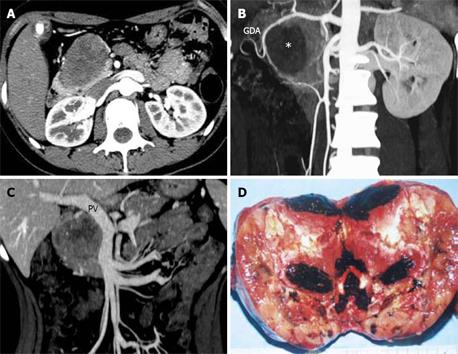
Figure 1 Pathologically confirmed benign SPT in a 24-year-old woman with abdominal discomfort for 1 year.
A: Axial image at arterial phase revealed a low-attenuation mass in the pancreatic head. The surrounding vessels were displaced; B: The MIP CTA image identified that the tumor (*) displaced the gastroduodenal artery (GDA) without infiltration; C: The MPVR image demonstrated that tumor compressed the portal vein (PV) with a smooth border. There was no evidence suggesting invasion; D: Hemorrhagic and cystic areas (dark areas) were detected in the gross specimen. The capsule was intact.
- Citation: Wang DB, Wang QB, Chai WM, Chen KM, Deng XX. Imaging features of solid pseudopapillary tumor of the pancreas on multi-detector row computed tomography. World J Gastroenterol 2009; 15(7): 829-835
- URL: https://www.wjgnet.com/1007-9327/full/v15/i7/829.htm
- DOI: https://dx.doi.org/10.3748/wjg.15.829


