Copyright
©2009 The WJG Press and Baishideng.
World J Gastroenterol. Feb 14, 2009; 15(6): 675-683
Published online Feb 14, 2009. doi: 10.3748/wjg.15.675
Published online Feb 14, 2009. doi: 10.3748/wjg.15.675
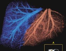
Figure 3 Forty-one-year-old male, potential living liver donor.
VR reconstruction shows normal anatomy of hepatic veins. The right lobe and right hepatic vein are blue, the left lobe and the MHV and left hepatic veins are red. A cut-plane runs 1 cm to the right of the MHV.
- Citation: Caruso S, Miraglia R, Maruzzelli L, Gruttadauria S, Luca A, Gridelli B. Imaging in liver transplantation. World J Gastroenterol 2009; 15(6): 675-683
- URL: https://www.wjgnet.com/1007-9327/full/v15/i6/675.htm
- DOI: https://dx.doi.org/10.3748/wjg.15.675


