Copyright
©2009 The WJG Press and Baishideng.
World J Gastroenterol. Feb 14, 2009; 15(6): 648-674
Published online Feb 14, 2009. doi: 10.3748/wjg.15.648
Published online Feb 14, 2009. doi: 10.3748/wjg.15.648
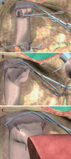
Figure 5 Anastomosis between the left hepatic vein of the graft and the inferior vena cava of the recipient, performed with the triangulation technique.
A: The bridge between the ostia of the right, middle, and left hepatic veins is cut to obtain a single opening; B: The opening is further enlarged by cutting the anterior face of the vena cava to obtain a wide triangular orifice; C: Three 5/0 vascular monofilament sutures are placed, taking the three corners of the graft and recipient orifices.
- Citation: Spada M, Riva S, Maggiore G, Cintorino D, Gridelli B. Pediatric liver transplantation. World J Gastroenterol 2009; 15(6): 648-674
- URL: https://www.wjgnet.com/1007-9327/full/v15/i6/648.htm
- DOI: https://dx.doi.org/10.3748/wjg.15.648


