Copyright
©2009 The WJG Press and Baishideng.
World J Gastroenterol. Feb 7, 2009; 15(5): 633-635
Published online Feb 7, 2009. doi: 10.3748/wjg.15.633
Published online Feb 7, 2009. doi: 10.3748/wjg.15.633
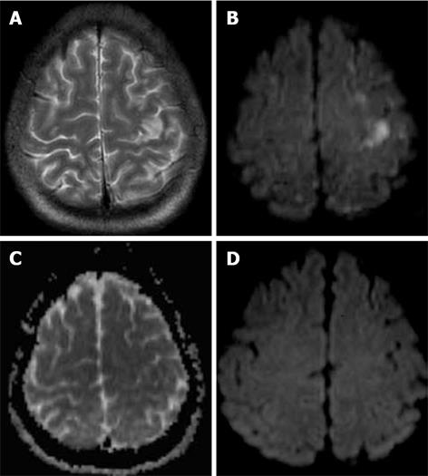
Figure 4 A 36-year-old man with cerebral lipiodol embolism.
A: Axial T2-weighted imaging (T2WI); B: Biffusion-weighted imaging (DWI); C: Apparent diffusion coefficient (ADC); D: Follow-up DWI. A, B and C obtained 57 h after the second TACE, and D obtained 5 wk after the second TACE. T2WI and DWI (A, B) demonstrated multiple high-signal lesions on the bilateral frontal and parietal lobe. The corresponding lesions showed a low signal area on the ADC map (C). A follow-up DWI revealed no obvious abnormal signal (D).
- Citation: Wu JJ, Chao M, Zhang GQ, Li B, Dong F. Pulmonary and cerebral lipiodol embolism after transcatheter arterial hemoembolization in hepatocellular carcinoma. World J Gastroenterol 2009; 15(5): 633-635
- URL: https://www.wjgnet.com/1007-9327/full/v15/i5/633.htm
- DOI: https://dx.doi.org/10.3748/wjg.15.633


