Copyright
©2009 The WJG Press and Baishideng.
World J Gastroenterol. Feb 7, 2009; 15(5): 628-632
Published online Feb 7, 2009. doi: 10.3748/wjg.15.628
Published online Feb 7, 2009. doi: 10.3748/wjg.15.628
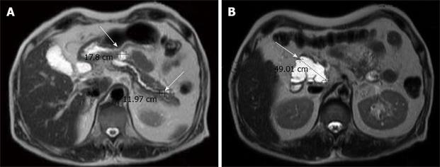
Figure 4 Magnetic resonance imaging showing three cystic lesions in the pancreatic head (4.
9 cm), body (1.8 cm) and tail (1.2 cm), with P-duct dilatation. Mild common bile duct and common hepatic duct dilatation without definite tumor was observed in the liver upon MRI.
- Citation: Chiang KC, Hsu JT, Chen HY, Jwo SC, Hwang TL, Jan YY, Yeh CN. Multifocal intraductal papillary mucinous neoplasm of the pancreas-A case report. World J Gastroenterol 2009; 15(5): 628-632
- URL: https://www.wjgnet.com/1007-9327/full/v15/i5/628.htm
- DOI: https://dx.doi.org/10.3748/wjg.15.628


