Copyright
©2009 The WJG Press and Baishideng.
World J Gastroenterol. Feb 7, 2009; 15(5): 622-627
Published online Feb 7, 2009. doi: 10.3748/wjg.15.622
Published online Feb 7, 2009. doi: 10.3748/wjg.15.622
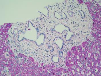
Figure 6 Histopathology of specimens taken from the hepatic parenchyma (PAS, × 200).
Distribution of many irregular, angulated duct structures was observed. Ductal epithelium was comprised of monolayered columnar epithelium, morphologically identical to biliary epithelium, surrounded by fibrous connective tissues with minimal inflammatory change. The sequential histopathological features were considered to be bile duct hamartoma (VMC). Subsequent immunohistochemistry of the region by anti-IgG4 antibody was negative.
- Citation: Miura H, Kitamura S, Yamada H. A variant form of autoimmune pancreatitis successfully treated by steroid therapy, accompanied by von Meyenburg complex. World J Gastroenterol 2009; 15(5): 622-627
- URL: https://www.wjgnet.com/1007-9327/full/v15/i5/622.htm
- DOI: https://dx.doi.org/10.3748/wjg.15.622


