Copyright
©2009 The WJG Press and Baishideng.
World J Gastroenterol. Feb 7, 2009; 15(5): 622-627
Published online Feb 7, 2009. doi: 10.3748/wjg.15.622
Published online Feb 7, 2009. doi: 10.3748/wjg.15.622
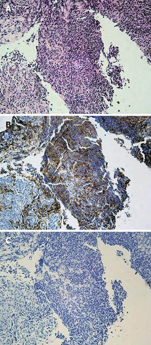
Figure 4 Histopathology of a specimen taken from the pancreatic head mass showed no malignant cells that indicated cancer.
A: Marked infiltration of pancreatic parenchyma by lymphocytic mononuclear cells, as well as interstitial fibrosis suggested a diagnosis of AIP (HE, × 200). B: These infiltrated cells were revealed to be mainly T lymphocytes (T-cell staining, × 200). However, the histopathology was not diagnostic without so-called LPSP with scarce plasma cell infiltration. C: Immunohistochmistry using antibody against IgG4 resulted in a negative study (IgG4 staining, × 200).
- Citation: Miura H, Kitamura S, Yamada H. A variant form of autoimmune pancreatitis successfully treated by steroid therapy, accompanied by von Meyenburg complex. World J Gastroenterol 2009; 15(5): 622-627
- URL: https://www.wjgnet.com/1007-9327/full/v15/i5/622.htm
- DOI: https://dx.doi.org/10.3748/wjg.15.622


