Copyright
©2009 The WJG Press and Baishideng.
World J Gastroenterol. Feb 7, 2009; 15(5): 622-627
Published online Feb 7, 2009. doi: 10.3748/wjg.15.622
Published online Feb 7, 2009. doi: 10.3748/wjg.15.622
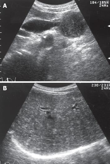
Figure 1 Abdominal US.
A: Pancreatic head mass with hypo-echogenicity, 43 mm in diameter, which compressed the lower portion of the bile duct, and resulted in upstream bile tract dilatation; B: Many tiny, diffuse hyperechoic patches or comet-like tails that suggested the presence of certain intrahepatic abnormalities, but the reason could not be elucidated.
- Citation: Miura H, Kitamura S, Yamada H. A variant form of autoimmune pancreatitis successfully treated by steroid therapy, accompanied by von Meyenburg complex. World J Gastroenterol 2009; 15(5): 622-627
- URL: https://www.wjgnet.com/1007-9327/full/v15/i5/622.htm
- DOI: https://dx.doi.org/10.3748/wjg.15.622


