Copyright
©2009 The WJG Press and Baishideng.
World J Gastroenterol. Feb 7, 2009; 15(5): 615-621
Published online Feb 7, 2009. doi: 10.3748/wjg.15.615
Published online Feb 7, 2009. doi: 10.3748/wjg.15.615
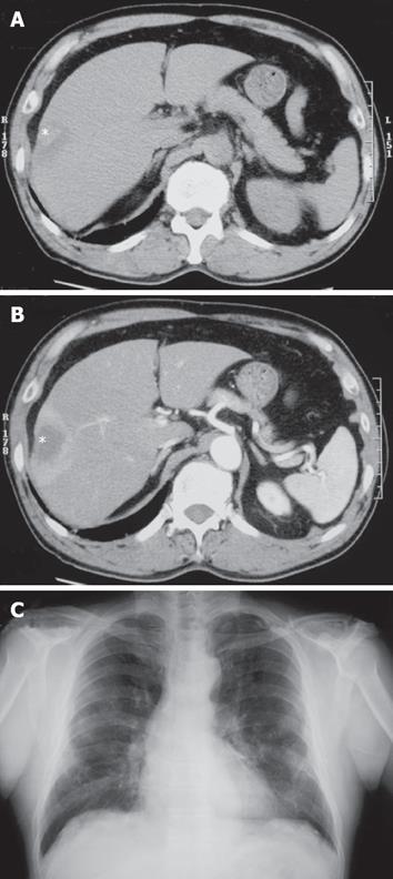
Figure 1 Radiological findings.
A: Plain CT showed a low-density mass (asterisk) in S8 segment of the liver; B: The periphery of the mass (asterisk) was enhanced in the early phase of enhanced CT, suggesting abundant blood supply; C: Chest X-ray showed pulmonary asbestosis and pleural thickening, but there were no new lesions suggesting lung cancer or pleural mesothelioma.
- Citation: Sasaki M, Araki I, Yasui T, Kinoshita M, Itatsu K, Nojima T, Nakanuma Y. Primary localized malignant biphasic mesothelioma of the liver in a patient with asbestosis. World J Gastroenterol 2009; 15(5): 615-621
- URL: https://www.wjgnet.com/1007-9327/full/v15/i5/615.htm
- DOI: https://dx.doi.org/10.3748/wjg.15.615


