Copyright
©2009 The WJG Press and Baishideng.
World J Gastroenterol. Feb 7, 2009; 15(5): 570-577
Published online Feb 7, 2009. doi: 10.3748/wjg.15.570
Published online Feb 7, 2009. doi: 10.3748/wjg.15.570
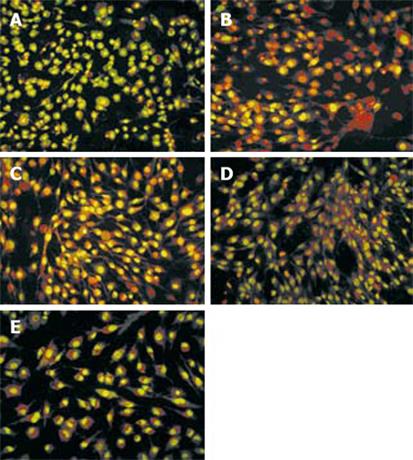
Figure 6 Apoptosis of mesothelial cells treated for 48 h.
A: serum free DMEM; B: MKN45; C: MKN45 + 200 &mgr;g/mL Astragalus injection; D: DMEM + 200 &mgr;g/mL Astragalus injection; E: GES-1 with AO/EB staining. Cells containing normal nuclear chromatin exhibit green nuclear staining. Cells containing fragmented nuclear chromatin exhibit orange to red nuclear staining.
- Citation: Na D, Liu FN, Miao ZF, Du ZM, Xu HM. Astragalus extract inhibits destruction of gastric cancer cells to mesothelial cells by anti-apoptosis. World J Gastroenterol 2009; 15(5): 570-577
- URL: https://www.wjgnet.com/1007-9327/full/v15/i5/570.htm
- DOI: https://dx.doi.org/10.3748/wjg.15.570


