Copyright
©2009 The WJG Press and Baishideng.
World J Gastroenterol. Dec 28, 2009; 15(48): 6044-6051
Published online Dec 28, 2009. doi: 10.3748/wjg.15.6044
Published online Dec 28, 2009. doi: 10.3748/wjg.15.6044
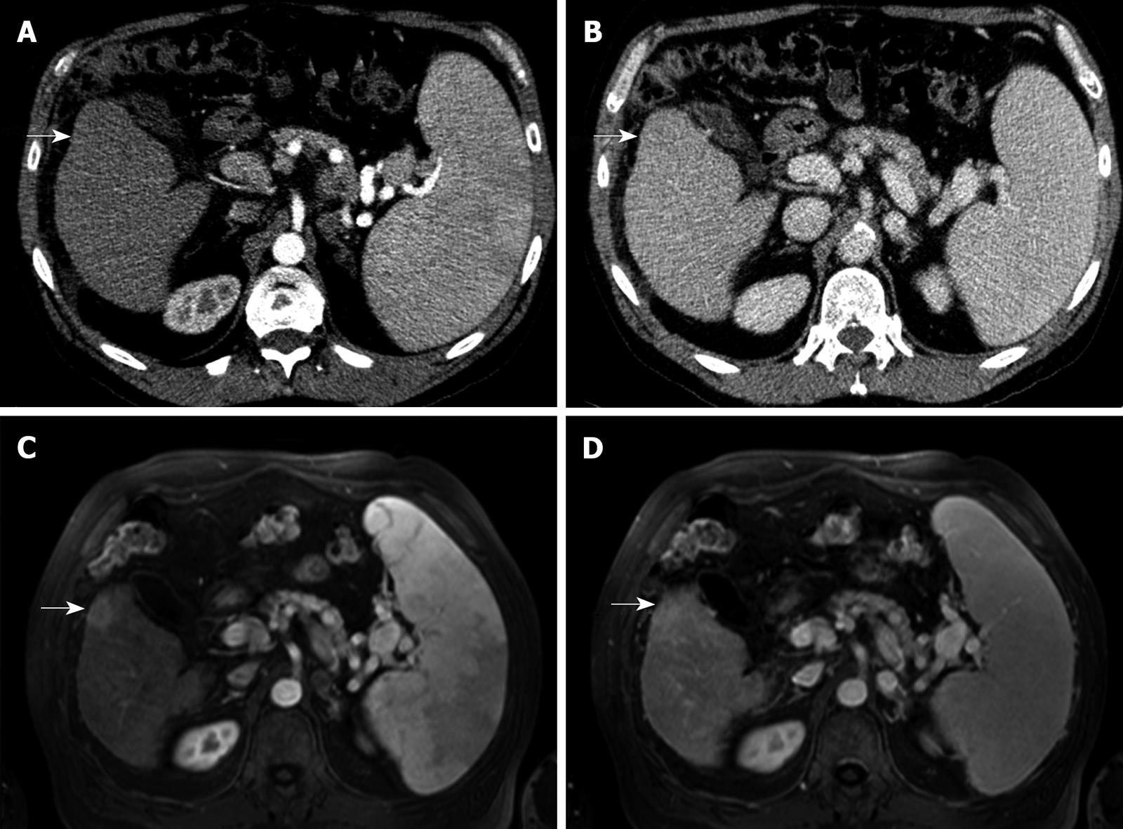
Figure 3 70-year-old man with biopsy-proven HCC.
Detection of an additional tumour nodule by MRI, size 19 mm (size category ≤ 20 mm). MDCT demonstrates no hypervascular enhancement in the contrast-enhanced arterial phase (A, arrow) or the portal venous phase (B, arrow). MRI arterial phase depicts a hypervascularized area in the T1w phase (C, arrow) which became isointense in the portal venous phase (D, arrow).
-
Citation: Pitton MB, Kloeckner R, Herber S, Otto G, Kreitner KF, Dueber C. MRI
versus 64-row MDCT for diagnosis of hepatocellular carcinoma. World J Gastroenterol 2009; 15(48): 6044-6051 - URL: https://www.wjgnet.com/1007-9327/full/v15/i48/6044.htm
- DOI: https://dx.doi.org/10.3748/wjg.15.6044


