Copyright
©2009 The WJG Press and Baishideng.
World J Gastroenterol. Dec 21, 2009; 15(47): 5898-5906
Published online Dec 21, 2009. doi: 10.3748/wjg.15.5898
Published online Dec 21, 2009. doi: 10.3748/wjg.15.5898
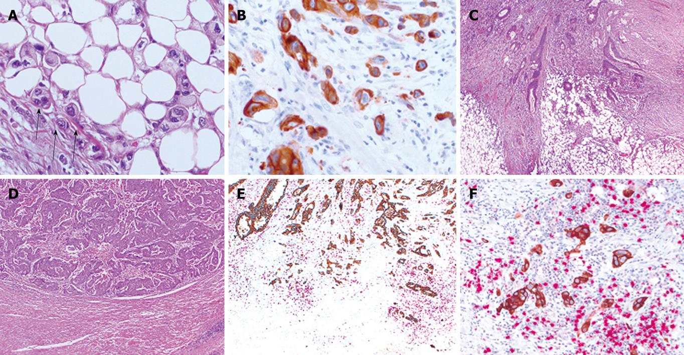
Figure 1 The invasive front of colorectal cancer.
A: HE staining of colorectal cancer (× 40 magnification) showing tumor buds (arrows) at the invasive front; B: Pan-cytokeratin staining (CK22) highlighting tumor buds at the invasive front of colorectal cancer (× 40 magnification). Colorectal cancers with different tumor border configurations upon evaluation of HE staining at low magnification (× 5); C: Infiltrating tumor border configuration; D: Pushing tumor border configuration; E: Low (× 5); F: High (× 20) power magnification of double immunostaining for pan-cytokeratin (CK22) and anti-CD8 antibody highlighting “attackers” (tumor buds, brown) and “defenders” (CD8+ T-lymphocytes, red) at the invasive front of a colorectal cancer with infiltrating tumor border configuration.
- Citation: Zlobec I, Lugli A. Invasive front of colorectal cancer: Dynamic interface of pro-/anti-tumor factors. World J Gastroenterol 2009; 15(47): 5898-5906
- URL: https://www.wjgnet.com/1007-9327/full/v15/i47/5898.htm
- DOI: https://dx.doi.org/10.3748/wjg.15.5898


