Copyright
©2009 The WJG Press and Baishideng.
World J Gastroenterol. Dec 21, 2009; 15(47): 5889-5897
Published online Dec 21, 2009. doi: 10.3748/wjg.15.5889
Published online Dec 21, 2009. doi: 10.3748/wjg.15.5889
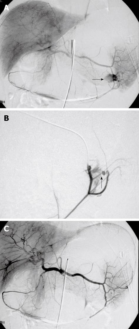
Figure 3 Digital subtraction images from a 37-year-old man with massive hematemesis.
A, B: Selective angiography shows a bleeding ulcer in the fundus of the stomach. Extravasation of contrast medium from a branch of the left gastroepiploic artery is seen (arrows); C: The control angiogram after glue embolization throughout the splenic artery shows complete and selective occlusion of the bleeding branch, with no active bleeding. The patient was discharged from the hospital 4 d later.
- Citation: Loffroy R, Guiu B. Role of transcatheter arterial embolization for massive bleeding from gastroduodenal ulcers. World J Gastroenterol 2009; 15(47): 5889-5897
- URL: https://www.wjgnet.com/1007-9327/full/v15/i47/5889.htm
- DOI: https://dx.doi.org/10.3748/wjg.15.5889


