Copyright
©2009 The WJG Press and Baishideng.
World J Gastroenterol. Dec 21, 2009; 15(47): 5889-5897
Published online Dec 21, 2009. doi: 10.3748/wjg.15.5889
Published online Dec 21, 2009. doi: 10.3748/wjg.15.5889
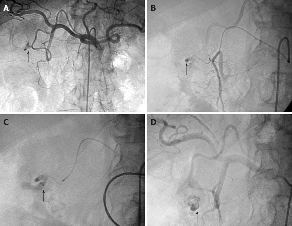
Figure 1 Arteriogram images of bleeding from a bulbar duodenal ulcer in a 76-year-old man.
A, B: Arteriogram showing contrast medium extravasated from a slender branch of the gastroduodenal artery (GDA) into the duodenum (arrows); C, D: After microcatheterization, selective glue embolization (radiopaque because of associated lipiodol (arrows) preserving the GDA ensured control of the bleeding, with no early or late recurrences.
- Citation: Loffroy R, Guiu B. Role of transcatheter arterial embolization for massive bleeding from gastroduodenal ulcers. World J Gastroenterol 2009; 15(47): 5889-5897
- URL: https://www.wjgnet.com/1007-9327/full/v15/i47/5889.htm
- DOI: https://dx.doi.org/10.3748/wjg.15.5889


