Copyright
©2009 The WJG Press and Baishideng.
World J Gastroenterol. Dec 14, 2009; 15(46): 5859-5863
Published online Dec 14, 2009. doi: 10.3748/wjg.15.5859
Published online Dec 14, 2009. doi: 10.3748/wjg.15.5859
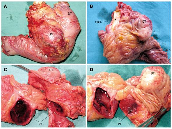
Figure 2 Resected duodenum and pancreatic head specimen containing both tumors.
A: Posterior aspect; B: Resected duodenum highlighting the duodenal tumor and common bile duct; C, D: Transected head of the pancreas showing the cystic pancreatic and duodenal tumors. DT: Duodenal tumor; PT: Pancreatic tumor; CBD: Common bile duct.
- Citation: Čolović RB, Matić SV, Micev MT, Grubor NM, Atkinson HD, Latinčić SM. Two synchronous somatostatinomas of the duodenum and pancreatic head in one patient. World J Gastroenterol 2009; 15(46): 5859-5863
- URL: https://www.wjgnet.com/1007-9327/full/v15/i46/5859.htm
- DOI: https://dx.doi.org/10.3748/wjg.15.5859


