Copyright
©2009 The WJG Press and Baishideng.
World J Gastroenterol. Dec 14, 2009; 15(46): 5833-5837
Published online Dec 14, 2009. doi: 10.3748/wjg.15.5833
Published online Dec 14, 2009. doi: 10.3748/wjg.15.5833
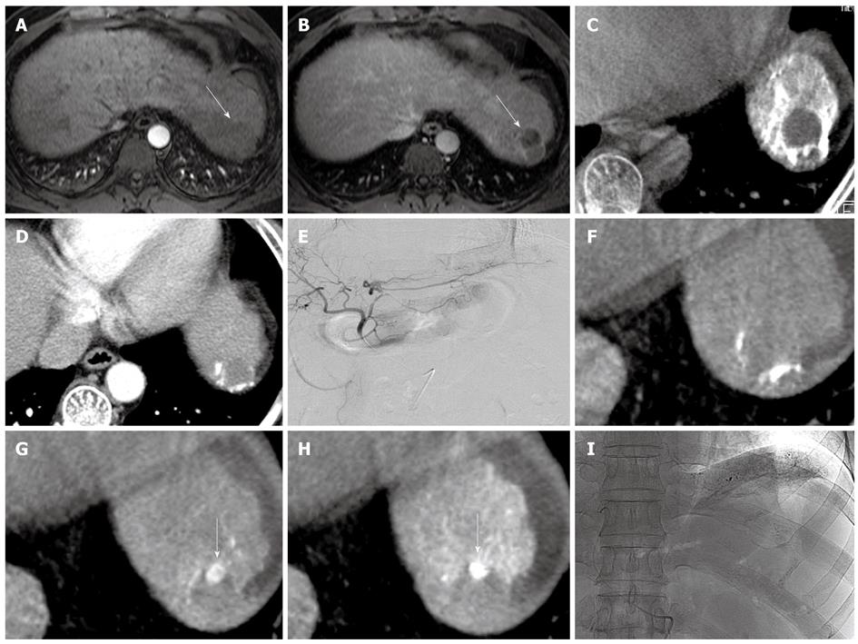
Figure 2 A 52-year-old man with HCC in S2 for 2nd TACE.
A, B: Early arterial phase (A) and delayed phase (B) of MRI reveals nodule (arrow) in segment 2 with no enhancement (arrow); C: Cone-beam CT after TACE shows poor grade of iodized oil uptake by the tumor; D: Early arterial phase of MDCT shows washout of iodized oil uptake near nodule, but no enhancing portion within the nodule 3 mo later; E: Left hepatic angiogram during TACE reveals no tumor and feeding artery 1 mo later; F, G: Cone-beam CT with (F) and without (G) hepatic arteriography shows small enhancing nodule (arrow) in the nodule; H, I: Cone-beam CT after infusion of emulsion (H) shows excellent grade iodized oil uptake by the nodule (arrow). This case shows typical “nodule-in-nodule” appearance of HCC, but spot image (I) did not distinguish nodular uptake.
- Citation: Jeon UB, Lee JW, Choo KS, Kim CW, Kim S, Lee TH, Jeong YJ, Kang DH. Iodized oil uptake assessment with cone-beam CT in chemoembolization of small hepatocellular carcinomas. World J Gastroenterol 2009; 15(46): 5833-5837
- URL: https://www.wjgnet.com/1007-9327/full/v15/i46/5833.htm
- DOI: https://dx.doi.org/10.3748/wjg.15.5833


