Copyright
©2009 The WJG Press and Baishideng.
World J Gastroenterol. Dec 14, 2009; 15(46): 5833-5837
Published online Dec 14, 2009. doi: 10.3748/wjg.15.5833
Published online Dec 14, 2009. doi: 10.3748/wjg.15.5833
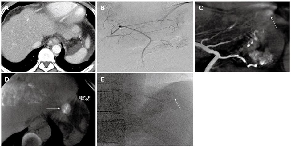
Figure 1 A 47-year-old man with hepatocellular carcinoma in S2 for 2nd TACE.
A: Preprocedural late arterial phase MDCT scan reveals hypervascular HCC near the diaphragm (arrow); B: Left gastric angiogram shows aberrant left hepatic artery with no tumor staining in suspected area; C: Tumor staining (arrow) and feeder artery are found after acquiring MIP image from cone-beam CT hepatic arteriography (Cone-beam CTHA); D: Cone-beam CT directly after TACE shows good grade iodized oil uptake in S2 (arrow); E: Spot image shows subtle lipiodol uptake near the left hemidiaphragm (arrow), but nodular iodized oil uptake is not observed.
- Citation: Jeon UB, Lee JW, Choo KS, Kim CW, Kim S, Lee TH, Jeong YJ, Kang DH. Iodized oil uptake assessment with cone-beam CT in chemoembolization of small hepatocellular carcinomas. World J Gastroenterol 2009; 15(46): 5833-5837
- URL: https://www.wjgnet.com/1007-9327/full/v15/i46/5833.htm
- DOI: https://dx.doi.org/10.3748/wjg.15.5833


