Copyright
©2009 The WJG Press and Baishideng.
World J Gastroenterol. Dec 7, 2009; 15(45): 5732-5739
Published online Dec 7, 2009. doi: 10.3748/wjg.15.5732
Published online Dec 7, 2009. doi: 10.3748/wjg.15.5732
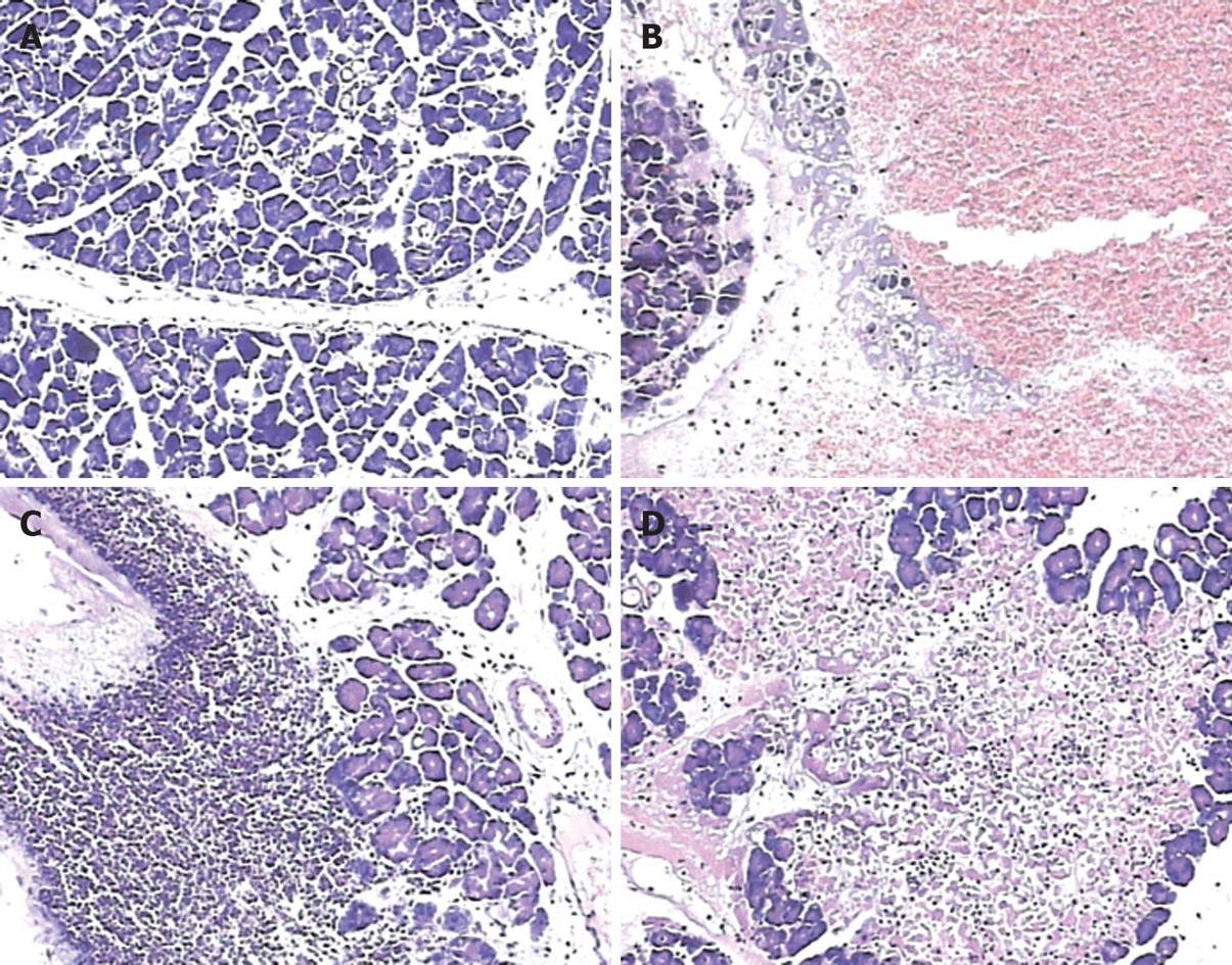
Figure 2 Pancreatic pathology (HE, × 100).
A: Normal pancreatic histology; B: Pancreatic damage induced 6 h after pancreatitis induction. Typical pathological features were edema and hemorrhage; C: Apparent leukocyte infiltration was observed 24 h after surgery; D: Pancreatic pathology induced 48 h after surgery, showing a confluent area of acinar cell necrosis.
- Citation: Liu ZH, Peng JS, Li CJ, Yang ZL, Xiang J, Song H, Wu XB, Chen JR, Diao DC. A simple taurocholate-induced model of severe acute pancreatitis in rats. World J Gastroenterol 2009; 15(45): 5732-5739
- URL: https://www.wjgnet.com/1007-9327/full/v15/i45/5732.htm
- DOI: https://dx.doi.org/10.3748/wjg.15.5732


