Copyright
©2009 The WJG Press and Baishideng.
World J Gastroenterol. Dec 7, 2009; 15(45): 5662-5668
Published online Dec 7, 2009. doi: 10.3748/wjg.15.5662
Published online Dec 7, 2009. doi: 10.3748/wjg.15.5662
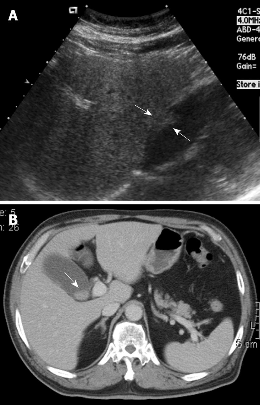
Figure 1 A 68-year-old woman with intermittent dull right upper quadrant pain for 6 mo.
A: Ultrasound (US) showed a sessile polypoid lesion (arrows) with a mildly uneven surface; B: Contrast-enhanced computed tomography (CT) showed an intraluminal polypoid lesion (arrow) with no apparent enlarged regional lymph node.
-
Citation: Lee TY, Ko SF, Huang CC, Ng SH, Liang JL, Huang HY, Chen MC, Sheen-Chen SM. Intraluminal
versus infiltrating gallbladder carcinoma: Clinical presentation, ultrasound and computed tomography. World J Gastroenterol 2009; 15(45): 5662-5668 - URL: https://www.wjgnet.com/1007-9327/full/v15/i45/5662.htm
- DOI: https://dx.doi.org/10.3748/wjg.15.5662


