Copyright
©2009 The WJG Press and Baishideng.
World J Gastroenterol. Nov 21, 2009; 15(43): 5493-5497
Published online Nov 21, 2009. doi: 10.3748/wjg.15.5493
Published online Nov 21, 2009. doi: 10.3748/wjg.15.5493
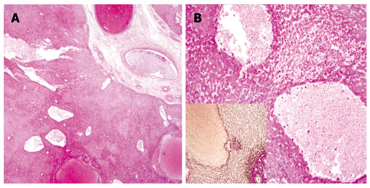
Figure 4 Microscopic findings.
A: Low magnification view shows variable-size, blood-filled cystic spaces (HE, × 10); B: High magnification view shows hemorrhagic necrosis in areas adjacent to peliotic spaces without lining endothelium (HE, × 100); Inset: immunohistochemical staining for reticulin (× 100).
- Citation: Choi SK, Jin JS, Cho SG, Choi SJ, Kim CS, Choe YM, Lee KY. Spontaneous liver rupture in a patient with peliosis hepatis: A case report. World J Gastroenterol 2009; 15(43): 5493-5497
- URL: https://www.wjgnet.com/1007-9327/full/v15/i43/5493.htm
- DOI: https://dx.doi.org/10.3748/wjg.15.5493


