Copyright
©2009 The WJG Press and Baishideng.
World J Gastroenterol. Nov 21, 2009; 15(43): 5489-5492
Published online Nov 21, 2009. doi: 10.3748/wjg.15.5489
Published online Nov 21, 2009. doi: 10.3748/wjg.15.5489
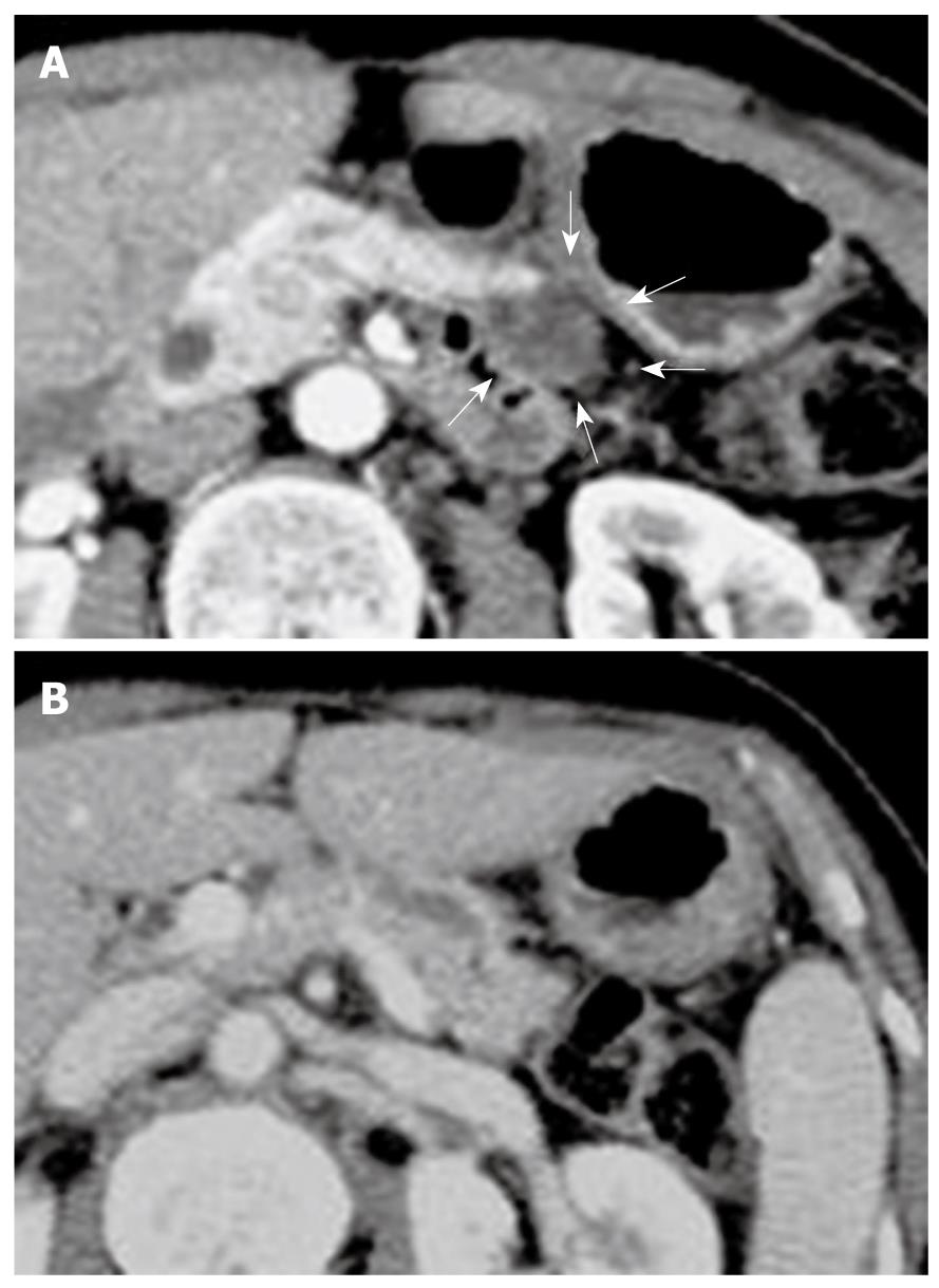
Figure 1 Contrast-enhanced computed tomography (CE-CT) scan of pancreas.
A: A cystic lesion in pancreatic body (arrows); B: A dilatation of the main duct in the pancreatic tail. Solid masses were not observed.
- Citation: Sakamoto H, Kitano M, Komaki T, Imai H, Kamata K, Kimura M, Takeyama Y, Kudo M. Small invasive ductal carcinoma of the pancreas distinct from branch duct intraductal papillary mucinous neoplasm. World J Gastroenterol 2009; 15(43): 5489-5492
- URL: https://www.wjgnet.com/1007-9327/full/v15/i43/5489.htm
- DOI: https://dx.doi.org/10.3748/wjg.15.5489


