Copyright
©2009 The WJG Press and Baishideng.
World J Gastroenterol. Nov 7, 2009; 15(41): 5236-5238
Published online Nov 7, 2009. doi: 10.3748/wjg.15.5236
Published online Nov 7, 2009. doi: 10.3748/wjg.15.5236
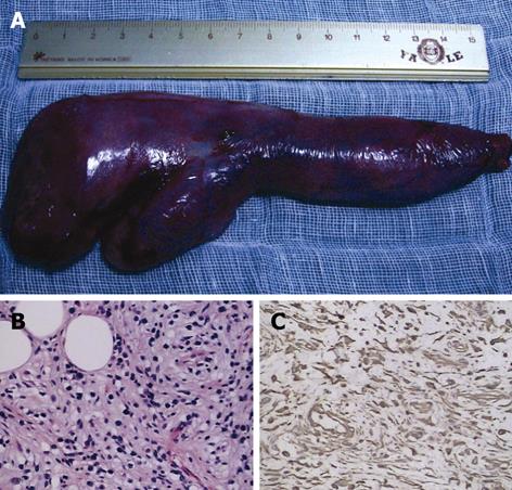
Figure 3 Polypectomy specimen and its pathology.
A: The resected esophageal lesion measured 17 cm in length, and the neck, body and tail of the lesion measured 1, 3 and 5 cm, respectively; B: HE staining (original magnification, × 200) showed the presence of lots of fibroblasts, with some acidophilic cells, plasma cells and adipose cells; C: Immunostaining (original magnification, × 200) for vimentin showed strong staining of the cells.
- Citation: Zhang J, Hao JY, Li SWH, Zhang ST. Successful endoscopic removal of a giant upper esophageal inflammatory fibrous polyp. World J Gastroenterol 2009; 15(41): 5236-5238
- URL: https://www.wjgnet.com/1007-9327/full/v15/i41/5236.htm
- DOI: https://dx.doi.org/10.3748/wjg.15.5236


