Copyright
©2009 The WJG Press and Baishideng.
World J Gastroenterol. Nov 7, 2009; 15(41): 5193-5199
Published online Nov 7, 2009. doi: 10.3748/wjg.15.5193
Published online Nov 7, 2009. doi: 10.3748/wjg.15.5193
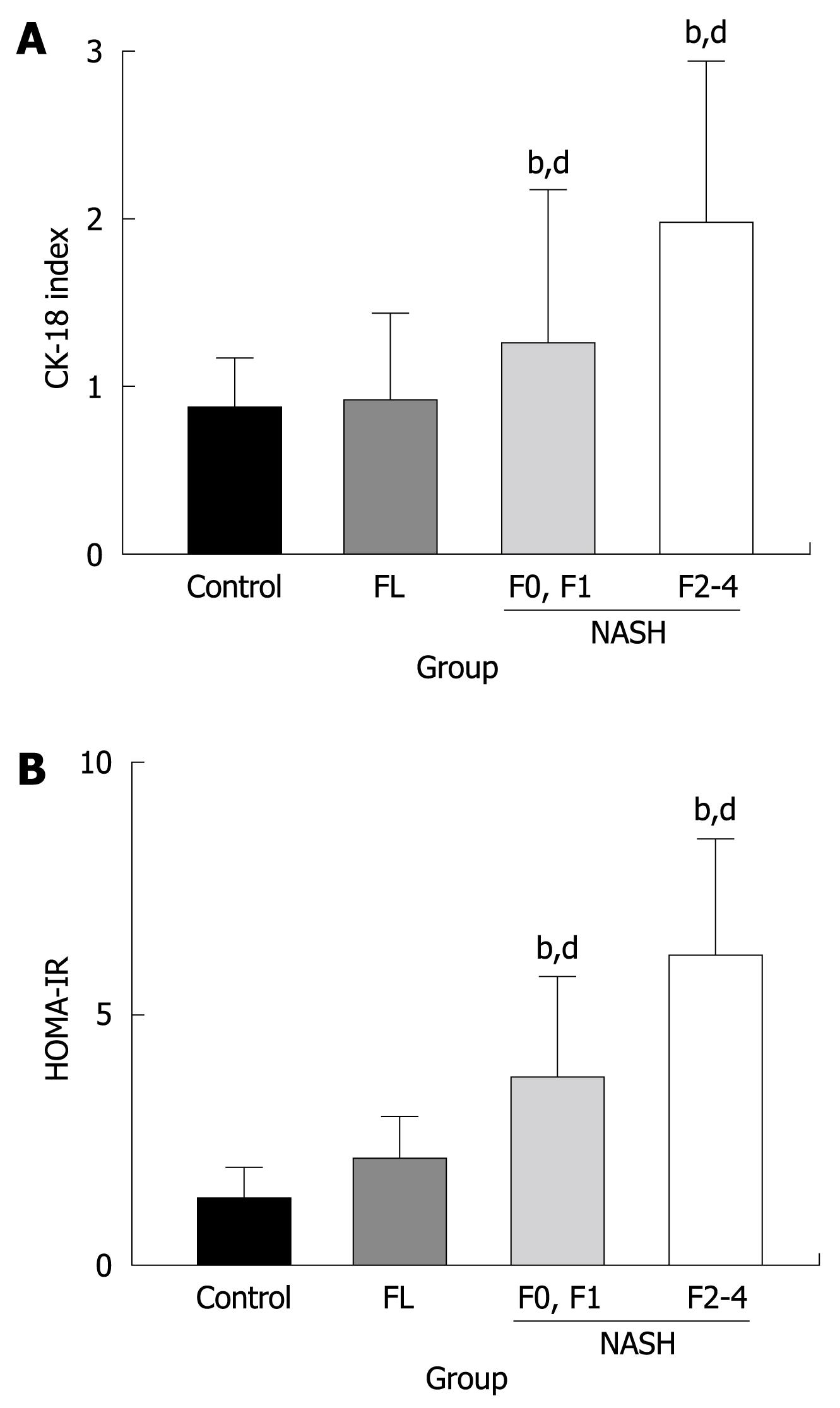
Figure 3 Semi-quantitative analysis of the CK-18-positive cells in the liver (A) and HOMA-IR (B) in NAFLD.
Similar to the CD34-positive neovascularization, there was no difference between the control healthy liver and FL. In NASH, a marked augmentation of CK-18 (A) and HOMA-IR (B) was found in the liver of NASH as compared with FL. The magnitude of neovascularization in high-grade (F2 to F4) liver fibrosis was more than in low-grade (F0, F1) fibrosis. The number of patients in each group was as follows; C (n = 3), FL (n = 11), low-grade fibrosis (F0 and F1: n = 10), and high-grade fibrosis (F2 to F4: n = 18). bP < 0.01 vs control group; dP < 0.01 vs low-grade fibrosis with NASH group.
- Citation: Kitade M, Yoshiji H, Noguchi R, Ikenaka Y, Kaji K, Shirai Y, Yamazaki M, Uemura M, Yamao J, Fujimoto M, Mitoro A, Toyohara M, Sawai M, Yoshida M, Morioka C, Tsujimoto T, Kawaratani H, Fukui H. Crosstalk between angiogenesis, cytokeratin-18, and insulin resistance in the progression of non-alcoholic steatohepatitis. World J Gastroenterol 2009; 15(41): 5193-5199
- URL: https://www.wjgnet.com/1007-9327/full/v15/i41/5193.htm
- DOI: https://dx.doi.org/10.3748/wjg.15.5193


