Copyright
©2009 The WJG Press and Baishideng.
World J Gastroenterol. Nov 7, 2009; 15(41): 5165-5175
Published online Nov 7, 2009. doi: 10.3748/wjg.15.5165
Published online Nov 7, 2009. doi: 10.3748/wjg.15.5165
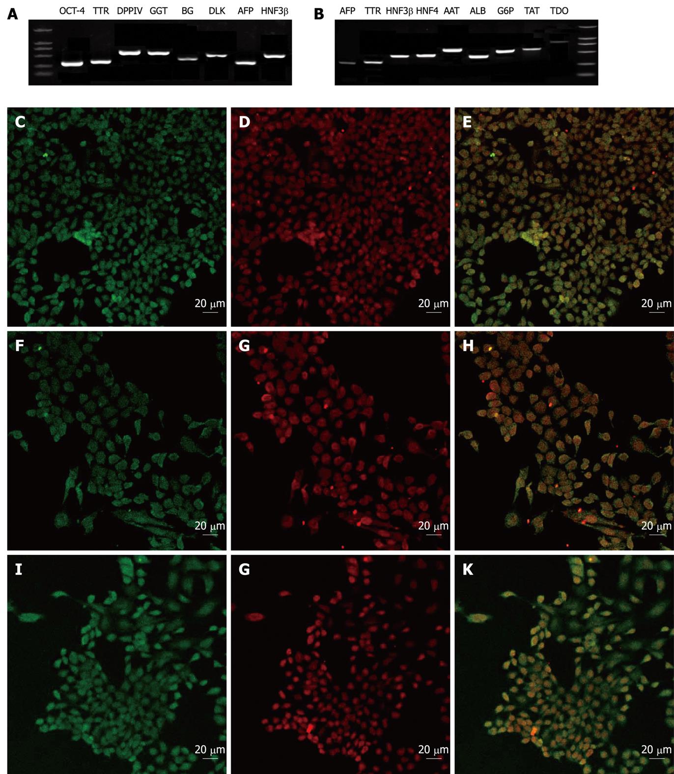
Figure 3 Gene expression analysis of VPA-induced hepatic lineage cells by RT-PCR and immunofluorescence staining.
A: RT-PCR showed hepatic progenitor cells after the treatment with valproic acid expressing most typical markers of hepatic/hepatic stem cells; B: The hepatic progenitor-derived hepatocytes expressing typical markers of mature liver cells; C-E: Immunofluorescent images of AFP (C) and OCT-4 (D) staining in VPA-induced hepatic progenitor cells (E is the merged image of C and D); F-H: Immunofluorescent images of AFP (F) and CK19 (G) staining in VPA-induced hepatic progenitor cells (H is the merged image of F and G); I-K: Immunofluorescent images of AFP (I) and DLK (J) staining in VPA-induced hepatic progenitor cells (K is the merged image of I and J).
- Citation: Dong XJ, Zhang GR, Zhou QJ, Pan RL, Chen Y, Xiang LX, Shao JZ. Direct hepatic differentiation of mouse embryonic stem cells induced by valproic acid and cytokines. World J Gastroenterol 2009; 15(41): 5165-5175
- URL: https://www.wjgnet.com/1007-9327/full/v15/i41/5165.htm
- DOI: https://dx.doi.org/10.3748/wjg.15.5165


