Copyright
©2009 The WJG Press and Baishideng.
World J Gastroenterol. Nov 7, 2009; 15(41): 5129-5140
Published online Nov 7, 2009. doi: 10.3748/wjg.15.5129
Published online Nov 7, 2009. doi: 10.3748/wjg.15.5129
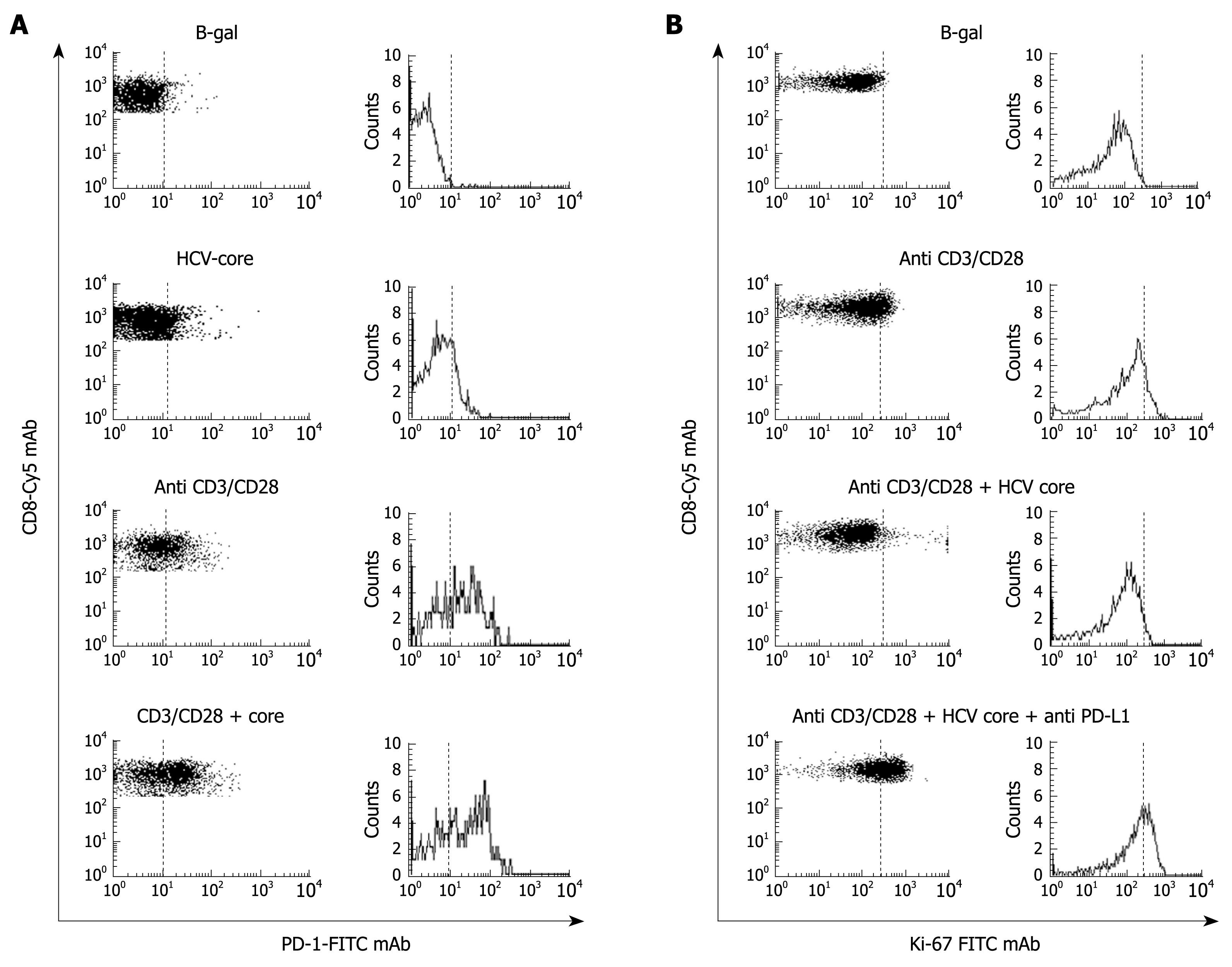
Figure 6 PD-1 up-regulation induced by HCV-core protein.
A: FACS® dot-plots and histograms of peripheral blood CD8+ cells stained with PD-1-FITC and CD8-Cy mAbs from a healthy subject. CD8+ cells were stimulated with B-galactosidase, HCV-core protein, CD3-CD28 mAb, and HCV-core protein plus CD3-CD28 mAbs. PD-1 expression was highly up-regulated on CD8+ cells after non-specific stimulation by anti CD3/CD28 mAbs in the presence of HCV core protein; B: FACS® dot-plots and histograms of peripheral blood CD8+ cells stained with Ki-67 FTIC and CD8-Cy mAbs from the same healthy subject to test proliferation ability of CD8+ cells after incubation with B-galactosidase, CD3-CD28 mAb, HCV-core protein plus CD3-CD28 mAb and HCV-core protein plus CD3-CD28 and anti-PD-L1 mAbs. HCV-core protein decreased the proliferation induced by CD3-CD28 mAb stimulation. This proliferation impairment induced by HCV-core protein was resolved by anti-PD-L1 mAb treatment.
- Citation: Larrubia JR, Benito-Martínez S, Miquel J, Calvino M, Sanz-de-Villalobos E, Parra-Cid T. Costimulatory molecule programmed death-1 in the cytotoxic response during chronic hepatitis C. World J Gastroenterol 2009; 15(41): 5129-5140
- URL: https://www.wjgnet.com/1007-9327/full/v15/i41/5129.htm
- DOI: https://dx.doi.org/10.3748/wjg.15.5129


