Copyright
©2009 The WJG Press and Baishideng.
World J Gastroenterol. Oct 21, 2009; 15(39): 4980-4983
Published online Oct 21, 2009. doi: 10.3748/wjg.15.4980
Published online Oct 21, 2009. doi: 10.3748/wjg.15.4980
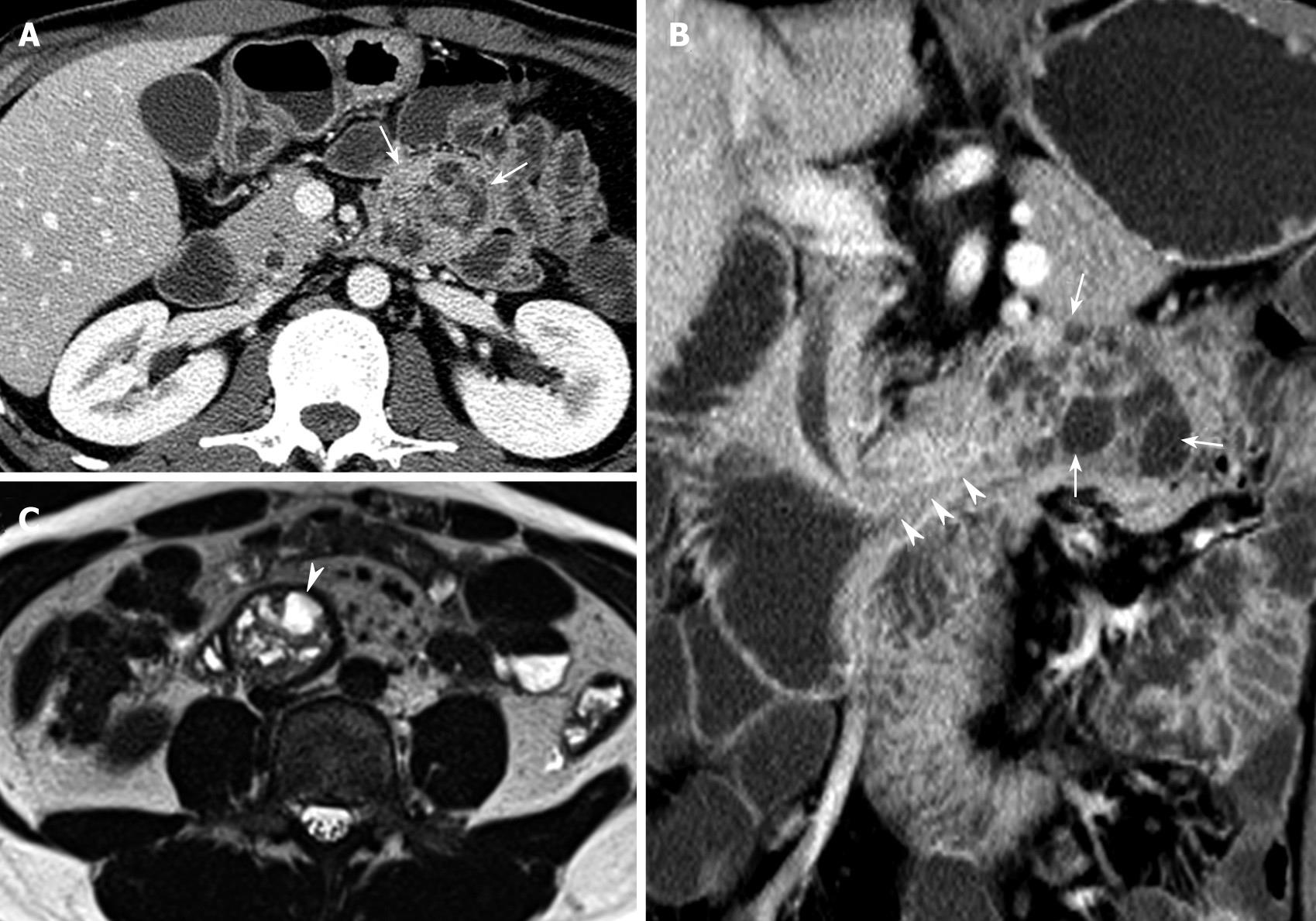
Figure 1 CT and MRI findings.
A: Axial portal venous-phase CT scan showing an intraluminal round mass (arrows) with internal multifocal low densities in the proximal jejunum; B: Coronal reformatted portal venous-phase CT demonstrating an identical configuration to the modified small bowel series showing a long stalk (arrowheads) and a well-defined mass with multifocal cystic lesions (arrows); C: A large, well-defined multi-chambered cystic mass showing high signal intensities (arrowhead) with its moved location in the third portion of the duodenum on axial T2-weighted images.
- Citation: Park BJ, Kim MJ, Lee JH, Park SS, Sung DJ, Cho SB. Cystic Brunner’s gland hamartoma in the duodenum: A case report. World J Gastroenterol 2009; 15(39): 4980-4983
- URL: https://www.wjgnet.com/1007-9327/full/v15/i39/4980.htm
- DOI: https://dx.doi.org/10.3748/wjg.15.4980


