Copyright
©2009 The WJG Press and Baishideng.
World J Gastroenterol. Oct 14, 2009; 15(38): 4856-4859
Published online Oct 14, 2009. doi: 10.3748/wjg.15.4856
Published online Oct 14, 2009. doi: 10.3748/wjg.15.4856
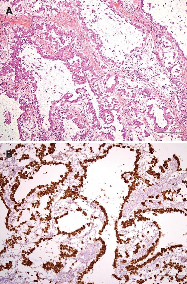
Figure 3 Photomicrographs of laparoscopically resected omental mass.
A: Proliferating epithelioid tumor cells formed tubular, cystic or papillary structures (HE, × 100, bar 100 μm); B: Tumor cells were strongly positive for calretinin upon immunohistochemical staining (× 100, bar 100 μm).
- Citation: Shin MK, Lee OJ, Ha CY, Min HJ, Kim TH. Malignant mesothelioma of the greater omentum mimicking omental infarction: A case report. World J Gastroenterol 2009; 15(38): 4856-4859
- URL: https://www.wjgnet.com/1007-9327/full/v15/i38/4856.htm
- DOI: https://dx.doi.org/10.3748/wjg.15.4856


