Copyright
©2009 The WJG Press and Baishideng.
World J Gastroenterol. Oct 14, 2009; 15(38): 4856-4859
Published online Oct 14, 2009. doi: 10.3748/wjg.15.4856
Published online Oct 14, 2009. doi: 10.3748/wjg.15.4856
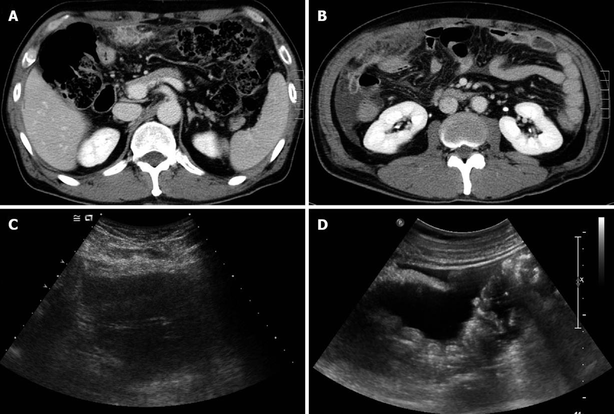
Figure 1 Abdominal computed tomography (CT) and ultrasonography (US).
A: Initial abdominal CT revealed a mass-like lesion of about 7 cm × 3 cm at the greater omentum; B: Abdominal CT follow-up after 1 wk antibiotic therapy showed an increased omental mass lesion, with a shift to the right lower abdomen, and ascites; C: Abdominal US showed a heterogeneous echoic mass at the greater omentum, and ascites; D: Multiple round nodules at the peritoneum and ascites.
- Citation: Shin MK, Lee OJ, Ha CY, Min HJ, Kim TH. Malignant mesothelioma of the greater omentum mimicking omental infarction: A case report. World J Gastroenterol 2009; 15(38): 4856-4859
- URL: https://www.wjgnet.com/1007-9327/full/v15/i38/4856.htm
- DOI: https://dx.doi.org/10.3748/wjg.15.4856


