Copyright
©2009 The WJG Press and Baishideng.
World J Gastroenterol. Oct 14, 2009; 15(38): 4816-4822
Published online Oct 14, 2009. doi: 10.3748/wjg.15.4816
Published online Oct 14, 2009. doi: 10.3748/wjg.15.4816
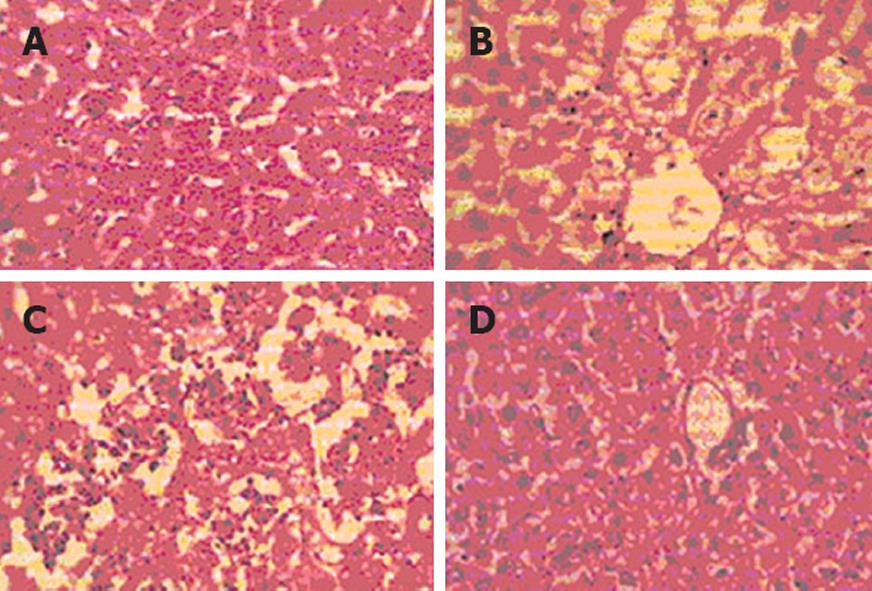
Figure 1 Photograph of rat liver (× 100).
A: Liver of a control rat showing normal hepatocytes and normal architecture; B: Liver section from a CCl4-treated rat demonstrating damaged liver cells and ballooning changes in the hepatocytes; C: Liver section from a Liv-52-treated rat showing mild feathery change, little ballooning degeneration of hepatocytes with associated normal hepatocytes; D: Liver section from a latifolia-treated rat showing a normal lobular pattern with minimal pooling of blood in the sinusoidal spaces.
-
Citation: Pradeep HA, Khan S, Ravikumar K, Ahmed MF, Rao MS, Kiranmai M, Reddy DS, Ahamed SR, Ibrahim M. Hepatoprotective evaluation of
Anogeissus latifolia :In vitro andin vivo studies. World J Gastroenterol 2009; 15(38): 4816-4822 - URL: https://www.wjgnet.com/1007-9327/full/v15/i38/4816.htm
- DOI: https://dx.doi.org/10.3748/wjg.15.4816


