Copyright
©2009 The WJG Press and Baishideng.
World J Gastroenterol. Oct 7, 2009; 15(37): 4695-4708
Published online Oct 7, 2009. doi: 10.3748/wjg.15.4695
Published online Oct 7, 2009. doi: 10.3748/wjg.15.4695
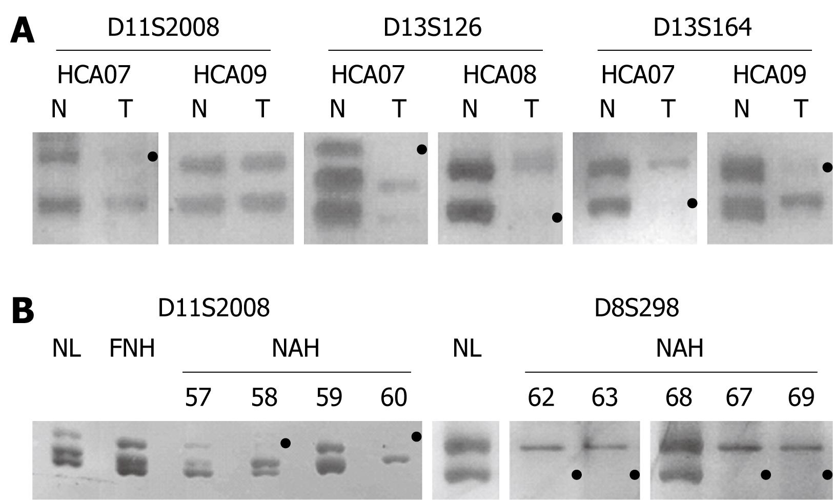
Figure 4 Representative data of amplification products at three length-polymorphic loci in HCAs 07-09 (A), FNH04 and NAH microdissected from the FNH lesion (B), with loss or marked reduction of the product from one allele (black dot) defined as LOH.
A: Gels show the allelic imbalance at D11S2008, D13S164 and D13S164 in HCA07, at D13S126 in HCA08, at D13S164, but not in D11S2008, in HCA09 (T: Tumor; N: Peritumorous normal liver); B: The microdissected lesions NAH58 and NAH60, but not NAH57 and NAH59, show LOH at D11S2008. The lesions NAH62, NAH63, NAH67 and NAH69, but not NAH68, show LOH at D8S298. Both alleles are preserved in FNH04 as tested as a whole (FNH) and compared to those of the surrounding normal liver parenchyma (NL).
- Citation: Cai YR, Gong L, Teng XY, Zhang HT, Wang CF, Wei GL, Guo L, Ding F, Liu ZH, Pan QJ, Su Q. Clonality and allelotype analyses of focal nodular hyperplasia compared with hepatocellular adenoma and carcinoma. World J Gastroenterol 2009; 15(37): 4695-4708
- URL: https://www.wjgnet.com/1007-9327/full/v15/i37/4695.htm
- DOI: https://dx.doi.org/10.3748/wjg.15.4695


