Copyright
©2009 The WJG Press and Baishideng.
World J Gastroenterol. Oct 7, 2009; 15(37): 4695-4708
Published online Oct 7, 2009. doi: 10.3748/wjg.15.4695
Published online Oct 7, 2009. doi: 10.3748/wjg.15.4695
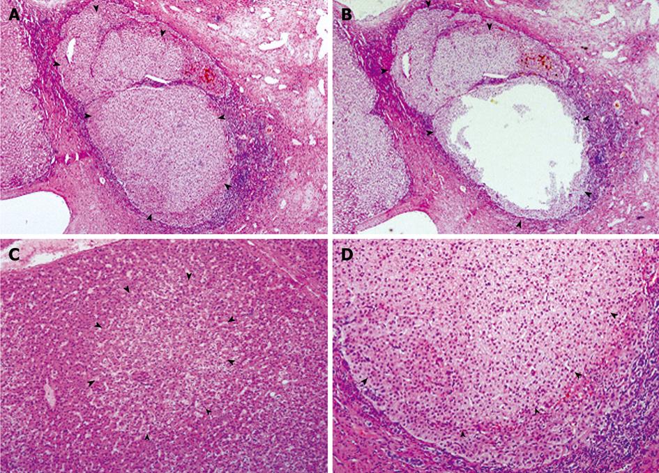
Figure 1 Preneoplastic lesions in classical FNH.
A and B: FNH04 from Case 03, showing a nodule largely occupied by an expanding NAH (arrowheads), before (A) and after microdissection (B); C: Portion of FNH06, showing an FAH (arrowheads), in which the altered hepatocytes integrate well with the surrounding hepatic plate; D: Portion of an NAH composed mainly of clear hepatocytes (arrowheads), showing compression to surrounding liver parenchyma. HE, A and B, × 40; C and D, × 100.
- Citation: Cai YR, Gong L, Teng XY, Zhang HT, Wang CF, Wei GL, Guo L, Ding F, Liu ZH, Pan QJ, Su Q. Clonality and allelotype analyses of focal nodular hyperplasia compared with hepatocellular adenoma and carcinoma. World J Gastroenterol 2009; 15(37): 4695-4708
- URL: https://www.wjgnet.com/1007-9327/full/v15/i37/4695.htm
- DOI: https://dx.doi.org/10.3748/wjg.15.4695


