Copyright
©2009 The WJG Press and Baishideng.
World J Gastroenterol. Sep 28, 2009; 15(36): 4587-4592
Published online Sep 28, 2009. doi: 10.3748/wjg.15.4587
Published online Sep 28, 2009. doi: 10.3748/wjg.15.4587
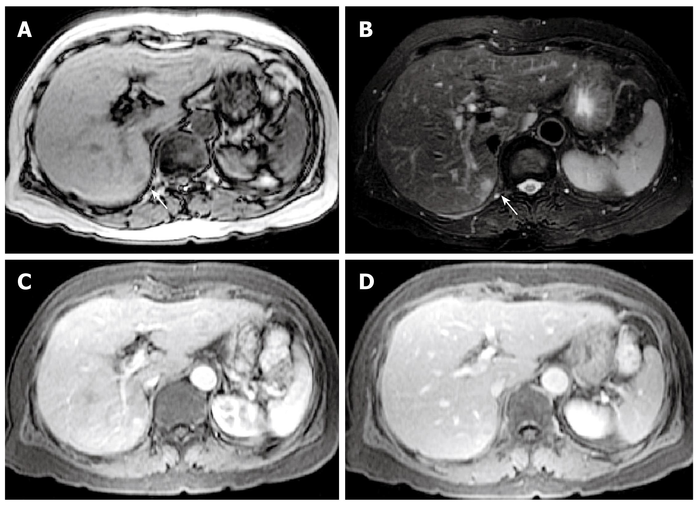
Figure 2 Magnetic resonance imaging (MRI) showing a 10 mm nodule in segment 7.
A: A hypointense nodule on T1-weighted images; B: A hyperintense nodule on T2-weighted images; C: A hyperintense nodule in the early phase after injection of contrast medium; D: A hypointense nodule in the late phase after injection of contrast medium.
- Citation: Okada T, Mibayashi H, Hasatani K, Hayashi Y, Tsuji S, Kaneko Y, Yoshimitsu M, Tani T, Zen Y, Yamagishi M. Pseudolymphoma of the liver associated with primary biliary cirrhosis: A case report and review of literature. World J Gastroenterol 2009; 15(36): 4587-4592
- URL: https://www.wjgnet.com/1007-9327/full/v15/i36/4587.htm
- DOI: https://dx.doi.org/10.3748/wjg.15.4587


