Copyright
©2009 The WJG Press and Baishideng.
World J Gastroenterol. Sep 28, 2009; 15(36): 4576-4581
Published online Sep 28, 2009. doi: 10.3748/wjg.15.4576
Published online Sep 28, 2009. doi: 10.3748/wjg.15.4576
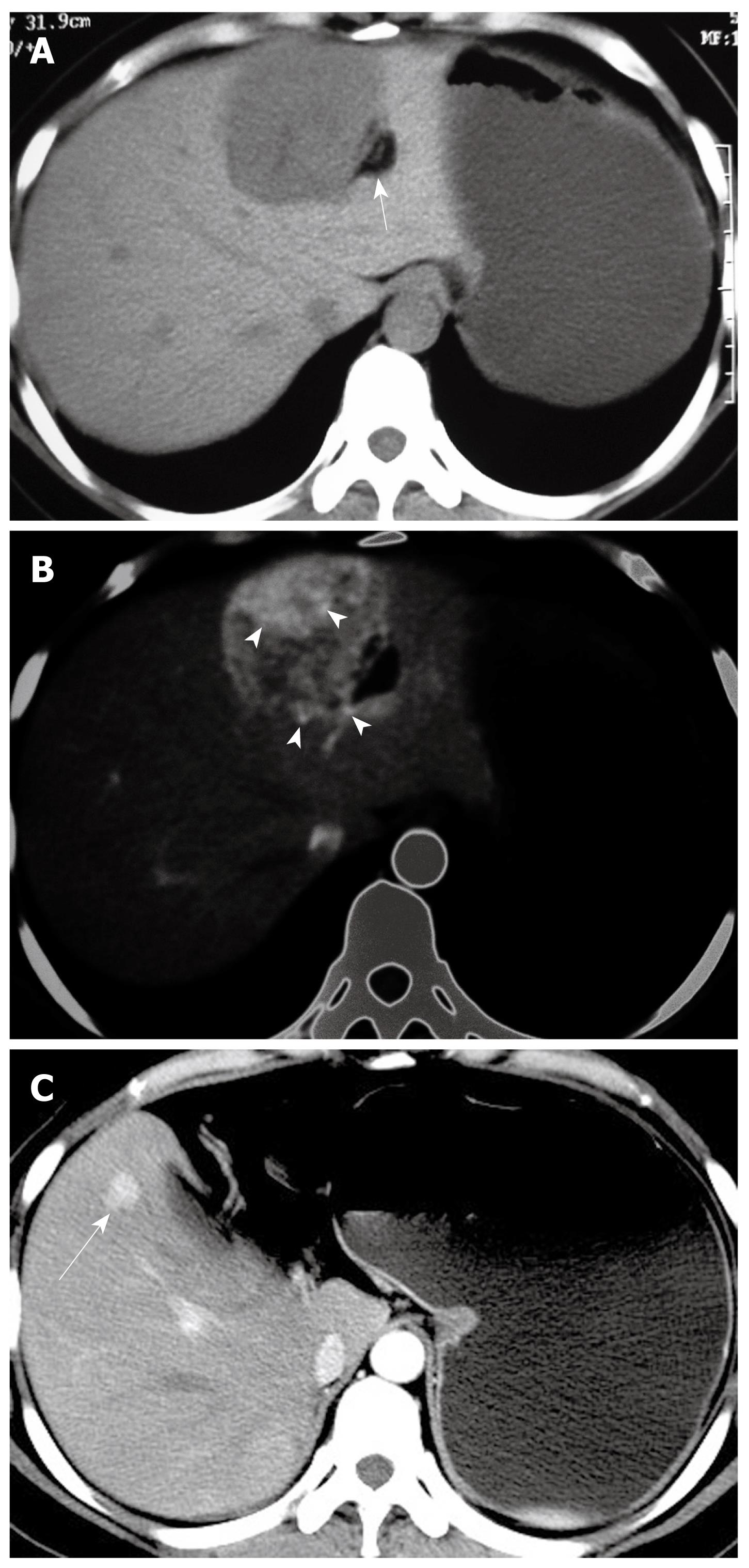
Figure 1 33-year-old woman with epithelioid angiomyolipoma in left lobe of liver (patient 7).
A: Non-enhanced CT scan shows hypoattenuating lesion with fat components (arrow, CT value mean-35 Hu) in segments II/III/IV; B: Contrast-enhanced CT scan shows inhomogeneous enhancement lesion with punctiform or filiform enhanced vessels (arrowheads, the window width and level was adjusted) on arterial phase; C: Contrast-enhanced CT scan at 1-year follow-up shows enhanced recurrent nodule (arrow) on arterial phase image after the left lobe surgery.
- Citation: Xu PJ, Shan Y, Yan FH, Ji Y, Ding Y, Zhou ML. Epithelioid angiomyolipoma of the liver: Cross-sectional imaging findings of 10 immunohistochemically-verified cases. World J Gastroenterol 2009; 15(36): 4576-4581
- URL: https://www.wjgnet.com/1007-9327/full/v15/i36/4576.htm
- DOI: https://dx.doi.org/10.3748/wjg.15.4576


