Copyright
©2009 The WJG Press and Baishideng.
World J Gastroenterol. Sep 21, 2009; 15(35): 4402-4409
Published online Sep 21, 2009. doi: 10.3748/wjg.15.4402
Published online Sep 21, 2009. doi: 10.3748/wjg.15.4402
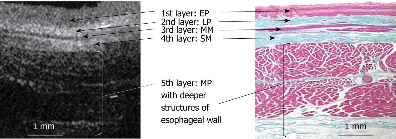
Figure 13 A schema of the correspondence between the OCT image of normal esophagus and histology.
The OCT image of normal esophageal wall delineates a five-layered morphology, consisting of relatively less reflective stratified squamous epithelium (the first layer), more reflective lamina propria (the second layer), less reflective muscularis mucosa (the third layer), more reflective submucosa (the fourth layer), and less reflective muscularis propria with deeper structures of esophageal wall (the fifth layer). (EM, original magnification × 4). The white and black bars represent 1 mm.
- Citation: Yokosawa S, Koike T, Kitagawa Y, Hatta W, Uno K, Abe Y, Iijima K, Imatani A, Ohara S, Shimosegawa T. Identification of the layered morphology of the esophageal wall by optical coherence tomography. World J Gastroenterol 2009; 15(35): 4402-4409
- URL: https://www.wjgnet.com/1007-9327/full/v15/i35/4402.htm
- DOI: https://dx.doi.org/10.3748/wjg.15.4402


