Copyright
©2009 The WJG Press and Baishideng.
World J Gastroenterol. Sep 21, 2009; 15(35): 4402-4409
Published online Sep 21, 2009. doi: 10.3748/wjg.15.4402
Published online Sep 21, 2009. doi: 10.3748/wjg.15.4402
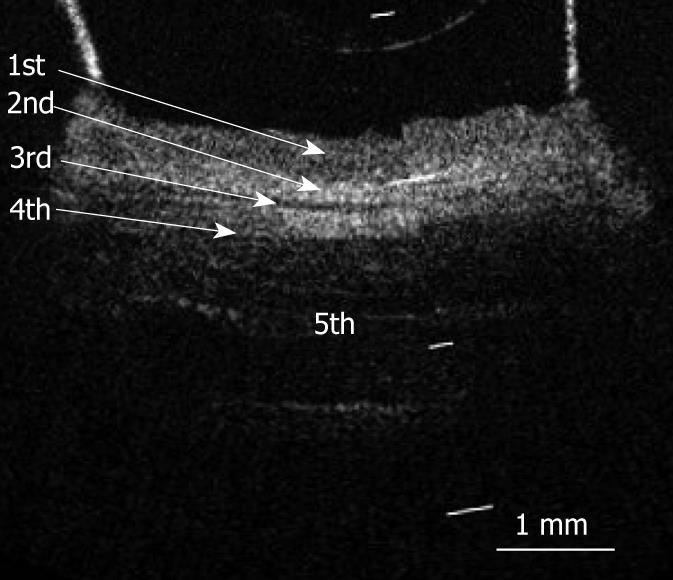
Figure 2 OCT image of a fresh pig esophageal wall specimen.
OCT image of the esophageal wall delineated a five-layered morphology (arrows) consisting of a relatively less reflective layer (the first layer), a more reflective layer (the second layer), a less reflective layer (the third layer), a more reflective layer (the fourth layer), and a less reflective layer (the fifth layer). The white bar represents 1 mm.
- Citation: Yokosawa S, Koike T, Kitagawa Y, Hatta W, Uno K, Abe Y, Iijima K, Imatani A, Ohara S, Shimosegawa T. Identification of the layered morphology of the esophageal wall by optical coherence tomography. World J Gastroenterol 2009; 15(35): 4402-4409
- URL: https://www.wjgnet.com/1007-9327/full/v15/i35/4402.htm
- DOI: https://dx.doi.org/10.3748/wjg.15.4402


