Copyright
©2009 The WJG Press and Baishideng.
World J Gastroenterol. Sep 21, 2009; 15(35): 4402-4409
Published online Sep 21, 2009. doi: 10.3748/wjg.15.4402
Published online Sep 21, 2009. doi: 10.3748/wjg.15.4402
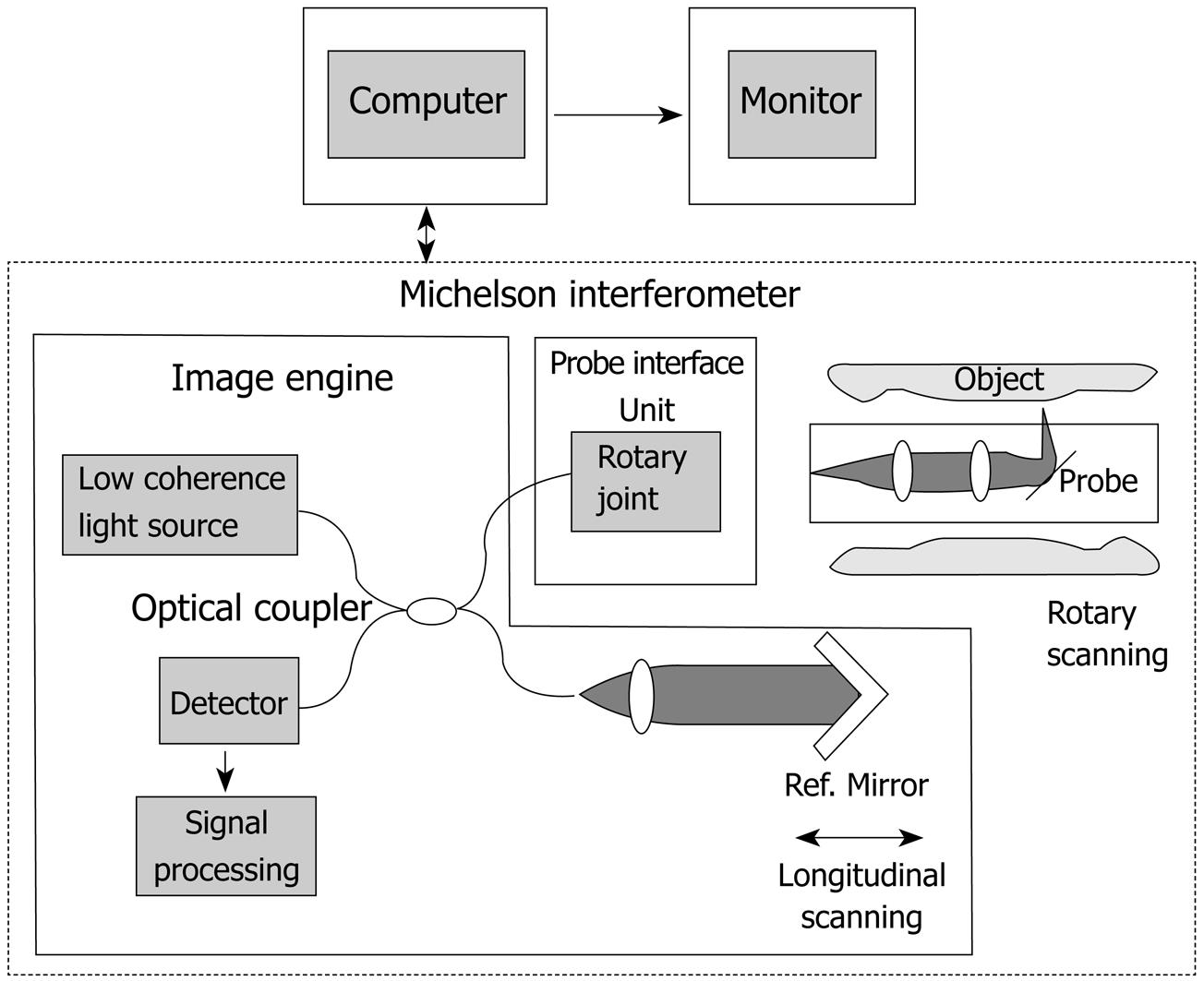
Figure 1 Schema of the OCT system (Light Lab Imaging, Boston, USA, and HOYA, Tokyo, Japan) used in the present study.
Near infrared light is generated from the light source, and then is split evenly. One beam is directed to the tissue sample and the other to a reference mirror. The light is reflected from both the sample and mirror. The reflected light beams are recombined in a beam splitter.
- Citation: Yokosawa S, Koike T, Kitagawa Y, Hatta W, Uno K, Abe Y, Iijima K, Imatani A, Ohara S, Shimosegawa T. Identification of the layered morphology of the esophageal wall by optical coherence tomography. World J Gastroenterol 2009; 15(35): 4402-4409
- URL: https://www.wjgnet.com/1007-9327/full/v15/i35/4402.htm
- DOI: https://dx.doi.org/10.3748/wjg.15.4402


