Copyright
©2009 The WJG Press and Baishideng.
World J Gastroenterol. Sep 14, 2009; 15(34): 4334-4338
Published online Sep 14, 2009. doi: 10.3748/wjg.15.4334
Published online Sep 14, 2009. doi: 10.3748/wjg.15.4334
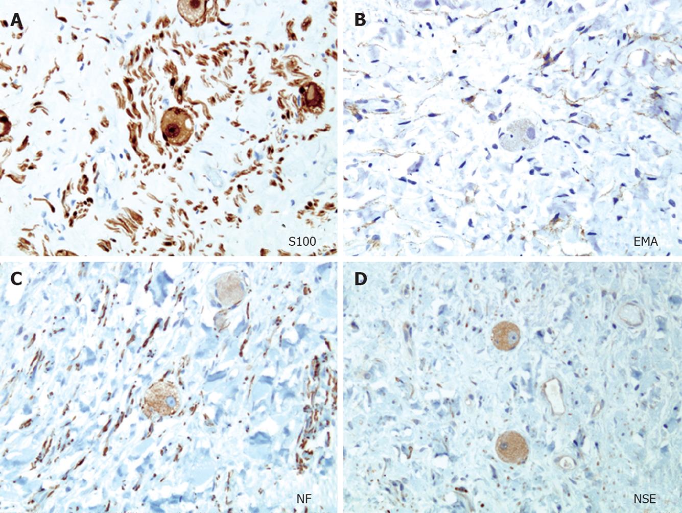
Figure 6 Representative immunocytochemistry samples of the typical ganglioneuroma (× 20).
A: S100 protein stain showing mostly Schwann cells and ganglion cells; B, C: Epithelial membrane antigen (EMA) and neurofilament (NF) stain showing the nerve fibers, highlighting the perineural cells and the axons of the nerve fibers; D: Ganglion cells are positive for neuronal-specific enolase (NSE).
- Citation: Poves I, Burdío F, Iglesias M, Martínez-Serrano MLÁ, Aguilar G, Grande L. Resection of the uncinate process of the pancreas due to a ganglioneuroma. World J Gastroenterol 2009; 15(34): 4334-4338
- URL: https://www.wjgnet.com/1007-9327/full/v15/i34/4334.htm
- DOI: https://dx.doi.org/10.3748/wjg.15.4334


