Copyright
©2009 The WJG Press and Baishideng.
World J Gastroenterol. Sep 14, 2009; 15(34): 4334-4338
Published online Sep 14, 2009. doi: 10.3748/wjg.15.4334
Published online Sep 14, 2009. doi: 10.3748/wjg.15.4334
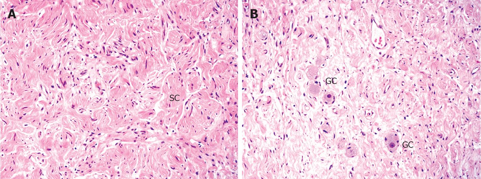
Figure 5 Microscopic images of the tumor (HE, × 10).
A: The lesion is composed of a proliferation of spindle-shaped cells in a whorled or fascicular pattern [Schwann cells (SC) and nerve fibers]. The cells have elongated and wavy nuclei with eosinophilic cytoplasm; B: Scattered mature ganglion cells (GC) are another of the histopathologic components of the tumor.
- Citation: Poves I, Burdío F, Iglesias M, Martínez-Serrano MLÁ, Aguilar G, Grande L. Resection of the uncinate process of the pancreas due to a ganglioneuroma. World J Gastroenterol 2009; 15(34): 4334-4338
- URL: https://www.wjgnet.com/1007-9327/full/v15/i34/4334.htm
- DOI: https://dx.doi.org/10.3748/wjg.15.4334


