Copyright
©2009 The WJG Press and Baishideng.
World J Gastroenterol. Sep 7, 2009; 15(33): 4204-4208
Published online Sep 7, 2009. doi: 10.3748/wjg.15.4204
Published online Sep 7, 2009. doi: 10.3748/wjg.15.4204
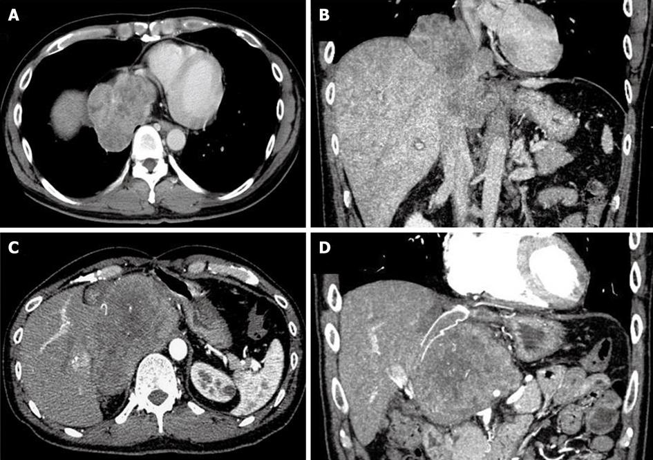
Figure 1 CT of the patient, showing the liver tumor infiltrating the suprahepatic inferior vena cava (A: Axial scan; B: Coronal scan), and showing the recurrent tumor (C: Axial scan; D: Coronal scan).
- Citation: Tomimaru Y, Nagano H, Marubashi S, Kobayashi S, Eguchi H, Takeda Y, Tanemura M, Kitagawa T, Umeshita K, Hashimoto N, Yoshikawa H, Wakasa K, Doki Y, Mori M. Sclerosing epithelioid fibrosarcoma of the liver infiltrating the inferior vena cava. World J Gastroenterol 2009; 15(33): 4204-4208
- URL: https://www.wjgnet.com/1007-9327/full/v15/i33/4204.htm
- DOI: https://dx.doi.org/10.3748/wjg.15.4204


