Copyright
©2009 The WJG Press and Baishideng.
World J Gastroenterol. Sep 7, 2009; 15(33): 4163-4169
Published online Sep 7, 2009. doi: 10.3748/wjg.15.4163
Published online Sep 7, 2009. doi: 10.3748/wjg.15.4163
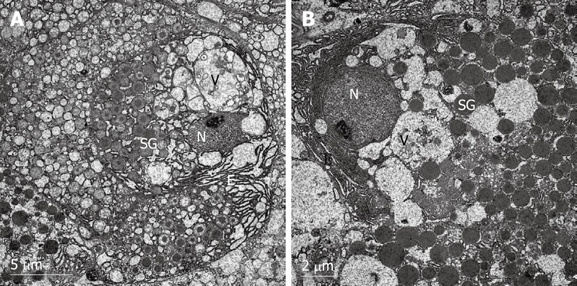
Figure 3 Electron micrographs of β-cells in rat pancreatic islets of control group.
N: Nucleus; SG: Secretory granule; V: Vesicle; E: Endoplasmic reticulum. The typical β-cells had an abundance of cytoplasmic granules and endoplasmic reticulum. The electron density of the granules increased when the granules were maturating in the vesicles. The mature secretory granules displayed a highly electron-dense core surrounded by a wide electron-lucent halo. The granules had a space between the core and the membrane. The vesicles showed a normal round outline (A, × 6000). There were many nascent granules in the β-cells; subsequent maturation involved further condensation of the matrix constituents and a reduction in granule diameter (B, × 8000).
- Citation: Qian TL, Wang XH, Liu S, Ma L, Lu Y. Fentanyl inhibits glucose-stimulated insulin release from β-cells in rat pancreatic islets. World J Gastroenterol 2009; 15(33): 4163-4169
- URL: https://www.wjgnet.com/1007-9327/full/v15/i33/4163.htm
- DOI: https://dx.doi.org/10.3748/wjg.15.4163


