Copyright
©2009 The WJG Press and Baishideng.
World J Gastroenterol. Sep 7, 2009; 15(33): 4143-4149
Published online Sep 7, 2009. doi: 10.3748/wjg.15.4143
Published online Sep 7, 2009. doi: 10.3748/wjg.15.4143
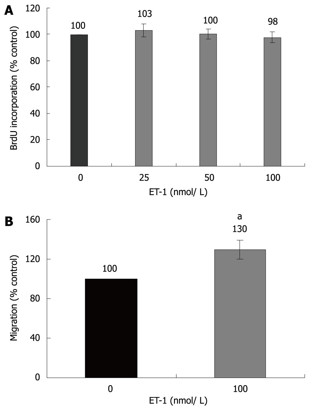
Figure 1 Effects of ET-1 on PSC proliferation and migration.
A: PSCs growing in 96-well plates were treated, under FCS-free conditions, with ET-1 at the indicated concentrations for 24 h. Cell proliferation was assessed with the BrdU DNA-incorporation assay. One hundred percent BrdU incorporation corresponds to untreated PSCs; B: CFSE-labelled cells were seeded into the upper chamber of 24-transwell plates, whereas ET-1 (100 nmol/L) was added to the lower chamber as indicated. Cell migration under FCS-free conditions was analyzed as described in the “Materials and Methods” section. One hundred percent cell migration corresponds to the intensity of the fluorescence signal received from untreated PSCs. Data in (A) and (B) are presented as mean ± SE (n≥ 6 separate cultures); aP < 0.05 vs control cultures.
- Citation: Jonitz A, Fitzner B, Jaster R. Molecular determinants of the profibrogenic effects of endothelin-1 in pancreatic stellate cells. World J Gastroenterol 2009; 15(33): 4143-4149
- URL: https://www.wjgnet.com/1007-9327/full/v15/i33/4143.htm
- DOI: https://dx.doi.org/10.3748/wjg.15.4143


