Case Report Open Access
Copyright ©2009 The WJG Press and Baishideng. All rights reserved.
World J Gastroenterol. Sep 7, 2009; 15(33): 4215-4217
Published online Sep 7, 2009. doi: 10.3748/wjg.15.4215
Enterovesical fistula caused by a bladder squamous cell carcinoma
Chun-Hsiang Ou Yang, Keng-Hao Liu, Phei-Lang Chang, Ta-Sen Yeh, Department of Surgery, Chang Gung Memorial Hospital, Chang Gung University, Taoyuan 333, Taiwan, China
Tse-Ching Chen, Department of Pathology, Chang Gung Memorial Hospital, Chang Gung University, Taoyuan 333, Taiwan, China
Author contributions: Ou Yang CH, and Liu KH contributed equally to this work. Ou Yang CH collected data and drafted the paper; Liu KH edited the paper; Yeh TS designed the research and reviewed a draft; Chen TC and Chang PL collected data.
Correspondence to: Ta-Sen Yeh, MD, PhD, Department of Surgery, Chang Gung Memorial Hospital, Chang Gung University, 5 Fu-Hsing Street, Kwei-Shan Shiang, Taoyuan 333, Taiwan, China. surglab@adm.cgmh.org.tw
Telephone: +886-3281200-3219 Fax: +886-3-3285818
Received: June 1, 2009
Revised: July 15, 2009
Accepted: July 22, 2009
Published online: September 7, 2009

Abstract

Enterovesical fistulas are not uncommon in patients with inflammatory or malignant colonic disease, however, fistulas secondary to primary bladder carcinomas are extremely rare. We herein reported a patient presenting with intractable urinary tract infection due to enterovesical fistula formation caused by a squamous cell carcinoma of the urinary bladder. This patient underwent en bloc resection of the bladder dome and involved ileum, and recovered uneventfully without urinary complaint. To the best of our knowledge, this is the first case reported in the literature.

Key Words: Enterovesical fistula; Squamous cell carcinoma; Urinary bladder; Malignant fistula


INTRODUCTION
Figure 1
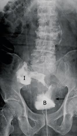
Figure 1 Cystography showed an enterovesical fistula (arrow). B: Urinary bladder; I: Ileum.
Figure 2
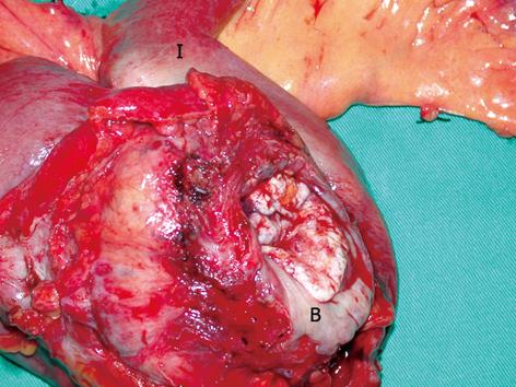
Figure 2 Surgical specimen of urinary bladder dome and ileum resected en bloc. B: Urinary bladder; I: Ileum.
Figure 3
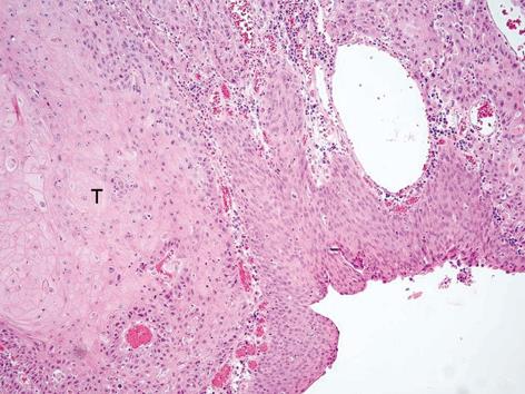
Figure 3 Microscopic examination yielded the diagnosis of squamous cell carcinoma of the urinary bladder. HE staining, original magnification × 40. T: Tumor burden.

Enterovesical fistulas are not uncommon in patients with inflammatory or malignant colonic disease, however, fistulas secondary to primary bladder carcinomas are extremely rare[1-3]. We herein report a patient presenting with intractable urinary tract infection due to enterovesical fistula formation caused by a squamous cell carcinoma of the urinary bladder. To the best of our knowledge, this is the first case reported in the literature.

CASE REPORT

An 83 year-old male presented with a 4-mo history of recurrent urinary tract infection. He had a history of benign prostate hyperplasia (American Urology Association score 24) that was under medical treatment. In order to correct urine retention and prevent urinary tract infection, photosensitive vaporization of the prostate had been performed. One month after the operation, the patient suffered from urinary tract infection again. The urine culture contained multiple flora of the gastro-intestinal tract including: Escherichia coli, Viridans streptococcus, Klebsiella, Pneumoniae and Enterococcus faecium. Retrograde urethrography showed leakage of the contrast and cystoscopy disclosed a fistula in the right posterior wall of the bladder with edematous tissue surrounding the fistula (Figure 1). Abdominal and pelvic computed tomography showed a very large neoplasm in the pelvic cavity with suspicion of vesicoileal fistula formation. Under the impression of malignant enterovesical fistula, the patient underwent surgical intervention. At laparotomy, a 10 cm-diameter whitish stony hard tumor was located in the pelvis, involving the distal ileum and urinary bladder, with a frank fistula formation. Resection of the urinary bladder dome and the involved ileum was performed in en bloc fashion (Figure 2). The pathology report yielded a moderately differentiated squamous cell carcinoma of the urinary bladder with direct invasion of the terminal ileum (Figure 3). This patient recovered uneventfully and was discharged without urinary tract infection on post-operative day 7.

DISCUSSION

Enterovesical fistula is a rare disease, with an estimated two to three patients per 10 000 hospital admissions, with annular incidence of 0.5 per 100 000[1]. In a literature review, 20% of vesicoenteral fistulas were rectovesical fistulas and 4%-5% were appendicovesical fistulas. Carson et al[2] reported 100 cases of enterovesical fistulas with 51% colonic diverticulitis, 16% colorectal cancer, 5% urinary bladder carcinoma and 16% due to other causes, including secondary to radiation necrosis, cervical cancer, tuberculoma and iatrogenic perforation.

The most common histologic type of bladder cancer leading to enterovesical fistula is transitional cell carcinoma. Primary squamous cell carcinoma of the bladder is a relatively rare tumor. The prevalence of squamous cell carcinoma varies depending on geographic location. It accounts for only 3%-7% of bladder cancers in the United States and 1% in England, but up to 75% in Egypt, where schistosomiasis is endemic[4]. Squamous cell carcinoma are usually related to chronic infection, bladder stones, chronic indwelling catheters or bladder diverticula. To our knowledge, this is the first report of vesicoenteral fistula as a result of bladder squamous cell carcinoma. Only one case of vesicorectal fistula from invasive rectal squamous cell carcinoma cancer had been reported[5]. Almost all squamous cell carcinomas are already advanced and muscle-infiltrative at the time of diagnosis[6]. Squamous cell carcinomas of the bladder have an unfavorable prognosis due to a local advanced stage at the time of presentation.

The most common clinical presentations of enterovesical fistulas are urinary tract infection (100%), pneumaturia (66%), fecaluria (50%), and hematuria (22.6%)[3].

Diagnostic tests include cystoscopy examination, retrograde cystography, and computed tomography. The diagnostic rate using cystoscopy examination is 77%-79%[2,7], while retrograde cystogram and computed tomography showed the fistulous tract in 66.6% and 83.3%, respectively[8,9]. Barium enema or small bowel series have a 20%-35% diagnostic rate to indicate the fistula[4]. Under cystoscopy examination, the fistula may be seen within a hypereremic area at an early stage of the disease, and with cystic mucosal hyperplasia or localized granulation tissue at the late stage[10,11]. Computed tomography has the advantage of showing the fistula tract or gas distinctly in the bladder. In addition, it is useful in delineating the extent of disease involvement, visualizing the anatomic relationship of the adjacent organs and detecting distant metastasis for malignant diseases, which accordingly allows tailoring of the management strategy[7,12,13].

Optimal work-up of a malignant of enterovesical fistula includes detection of the fistula and anatomic extension using the aforementioned diagnostic tests and definitive pathological confirmation, if possible. Except for elderly people in generally poor condition, diverting enterostomy and indwelling urethral catheters are not recommended as permanent treatments for enterovesical fistulae[3].

Whitely and Grabtald proposed that a one-stage en bloc resection of the colonic malignancy and involved bladder portion is a reasonable and safe procedure[7,11], avoiding a total cystectomy. Patients must be well-prepared, non-obstructive and without systemic infection before the surgery[1,11,12,14,15]. However, if resection of the bladder is too wide, resulting in a total cystectomy, reconstruction to restore intestinal continuity and provide adequate bladder capacity and continence is pertinent. On the other hand, if the tumor is deemed unresectable and there is a short life expectancy, a palliative surgical procedure with enterostomy, with or without urinary diversion, is suggested[12,16].

The 5-year survival rate for colon cancer with enterovesical fistula is 81% in nodal negative patients (T4N0), compared with 27% in nodal positive patients (T4N1)[17]. Looser et al[16] also suggested that colonic cancer with a perforation into the bladder could be treated by resection for a curative purpose, obtaining long-term survival in 50% of the patients[7,16]. Nevertheless, Vidal Sans et al[3] reported that surgical morbidity and mortality is relatively high, especially in fistula resulting from malignancy. Despite various methods of treatment for enterovesical fistula, the prognosis for patients with an advanced stage of malignant fistulae is dismal.

Footnotes

Peer reviewers: Gianlorenzo Dionigi, MD, FACS, ESES, ETA, Asscociate Professor of Surgery, Department of Surgical Sciences, University of Insubria, Ospedate di Circolo, v. guicciardini, 21100 Varese, Italy; Ian C Roberts-Thomson, Professor, Department of Gastroenterology and Hepatology, The Queen Elizabeth Hospital, 28 Woodville Road, Woodville South 5011, Australia

S- Editor Tian L L- Editor Lutze M E- Editor Ma WH

References
1.  Karamchandani MC, West CF Jr. Vesicoenteric fistulas. Am J Surg. 1984;147:681-683.  [PubMed]  [DOI]  [Cited in This Article: ]
2.  Carson CC, Malek RS, Remine WH. Urologic aspects of vesicoenteric fistulas. J Urol. 1978;119:744-746.  [PubMed]  [DOI]  [Cited in This Article: ]
3.  Vidal Sans J, Pradell Teigell J, Palou Redorta J, Villagrasa Serrano M, Banús Gassol JM. Review of 31 vesicointestinal fistulas: diagnosis and management. Eur Urol. 1986;12:21-27.  [PubMed]  [DOI]  [Cited in This Article: ]
4.  Messing EM, Walsh PC, Retik AB. Urothelial tumors of the urinary tract. Campbells urology. Philadelphia, Pa: Elsevier Science 2002; 2732-2765.  [PubMed]  [DOI]  [Cited in This Article: ]
5.  Kodama K, Mizuno T, Imahori T, Ida M, Matsubara F. Concurrent diagnosis of urothelial carcinoma and squamous cell carcinoma of the bladder in a patient with a vesicorectal fistula from invasive rectal cancer. Int J Urol. 2006;13:296-298.  [PubMed]  [DOI]  [Cited in This Article: ]
6.  Shaaban AA, Orkubi SA, Said MT, Yousef B, Abomelha MS. Squamous cell carcinoma of the urinary bladder. Ann Saudi Med. 1997;17:115-119.  [PubMed]  [DOI]  [Cited in This Article: ]
7.  Dawam D, Patel S, Kouriefs C, Masood S, Khan O, Sheriff MK. A "urological" enterovesical fistula. J Urol. 2004;172:943-944.  [PubMed]  [DOI]  [Cited in This Article: ]
8.  Goldman SM, Fishman EK, Gatewood OM, Jones B, Siegelman SS. CT in the diagnosis of enterovesical fistulae. AJR Am J Roentgenol. 1985;144:1229-1233.  [PubMed]  [DOI]  [Cited in This Article: ]
9.  Sarr MG, Fishman EK, Goldman SM, Siegelman SS, Cameron JL. Enterovesical fistula. Surg Gynecol Obstet. 1987;164:41-48.  [PubMed]  [DOI]  [Cited in This Article: ]
10.  Best JW, Davis RM. Vesicointestinal fistulas. J Urol. 1969;101:62-65.  [PubMed]  [DOI]  [Cited in This Article: ]
11.  Larsen A, Bjerklund Johansen TE, Solheim BM, Urnes T. Diagnosis and treatment of enterovesical fistula. Eur Urol. 1996;29:318-321.  [PubMed]  [DOI]  [Cited in This Article: ]
12.  Holmes SA, Christmas TJ, Kirby RS, Hendry WF. Management of colovesical fistulae associated with pelvic malignancy. Br J Surg. 1992;79:432-434.  [PubMed]  [DOI]  [Cited in This Article: ]
13.  Fretz PC, Kirby PA, O'Donnell MA. Intravesical colonic pseudotumor. J Urol. 2004;171:340.  [PubMed]  [DOI]  [Cited in This Article: ]
14.  Suits GS, Knoepp LF. A community experience with entericovesical fistulas. Am Surg. 1985;51:523-528.  [PubMed]  [DOI]  [Cited in This Article: ]
15.  Nemer FD, Sweetser TH Jr, Goldberg SM, Balcos EG, Schlottler JL, Christenson CE. How to manage colovesical fistula. Geriatrics. 1978;33:86-87.  [PubMed]  [DOI]  [Cited in This Article: ]
16.  Looser KG, Quan SH, Clark DG. Colo-urinary-tract fistula in the cancer patient. Dis Colon Rectum. 1979;22:143-148.  [PubMed]  [DOI]  [Cited in This Article: ]
17.  Whiteley HW Jr, Grabstald H. Conservative management of distal large bowel cancer invading the urinary bladder. Clin Bull. 1975;5:99-101.  [PubMed]  [DOI]  [Cited in This Article: ]