Copyright
©2009 The WJG Press and Baishideng.
World J Gastroenterol. Aug 28, 2009; 15(32): 4049-4054
Published online Aug 28, 2009. doi: 10.3748/wjg.15.4049
Published online Aug 28, 2009. doi: 10.3748/wjg.15.4049
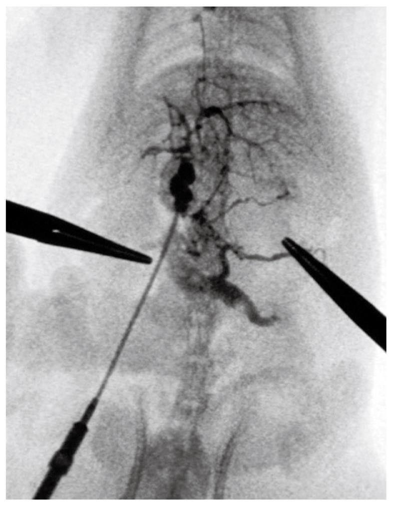
Figure 5 At week 10, portal venography showed the mesenteric vein with varices and collaterals.
A lot of contrast medium was seen in the vena cava, which indicated the establishment of the collateral circulation. These observations were not seen in the control rats. The left adrenal vein after PVL was clearly shown with an enlarged diameter, while this vein was not seen in the control group.
- Citation: Wen Z, Zhang JZ, Xia HM, Yang CX, Chen YJ. Stability of a rat model of prehepatic portal hypertension caused by partial ligation of the portal vein. World J Gastroenterol 2009; 15(32): 4049-4054
- URL: https://www.wjgnet.com/1007-9327/full/v15/i32/4049.htm
- DOI: https://dx.doi.org/10.3748/wjg.15.4049


