Copyright
©2009 The WJG Press and Baishideng.
World J Gastroenterol. Aug 28, 2009; 15(32): 4037-4043
Published online Aug 28, 2009. doi: 10.3748/wjg.15.4037
Published online Aug 28, 2009. doi: 10.3748/wjg.15.4037
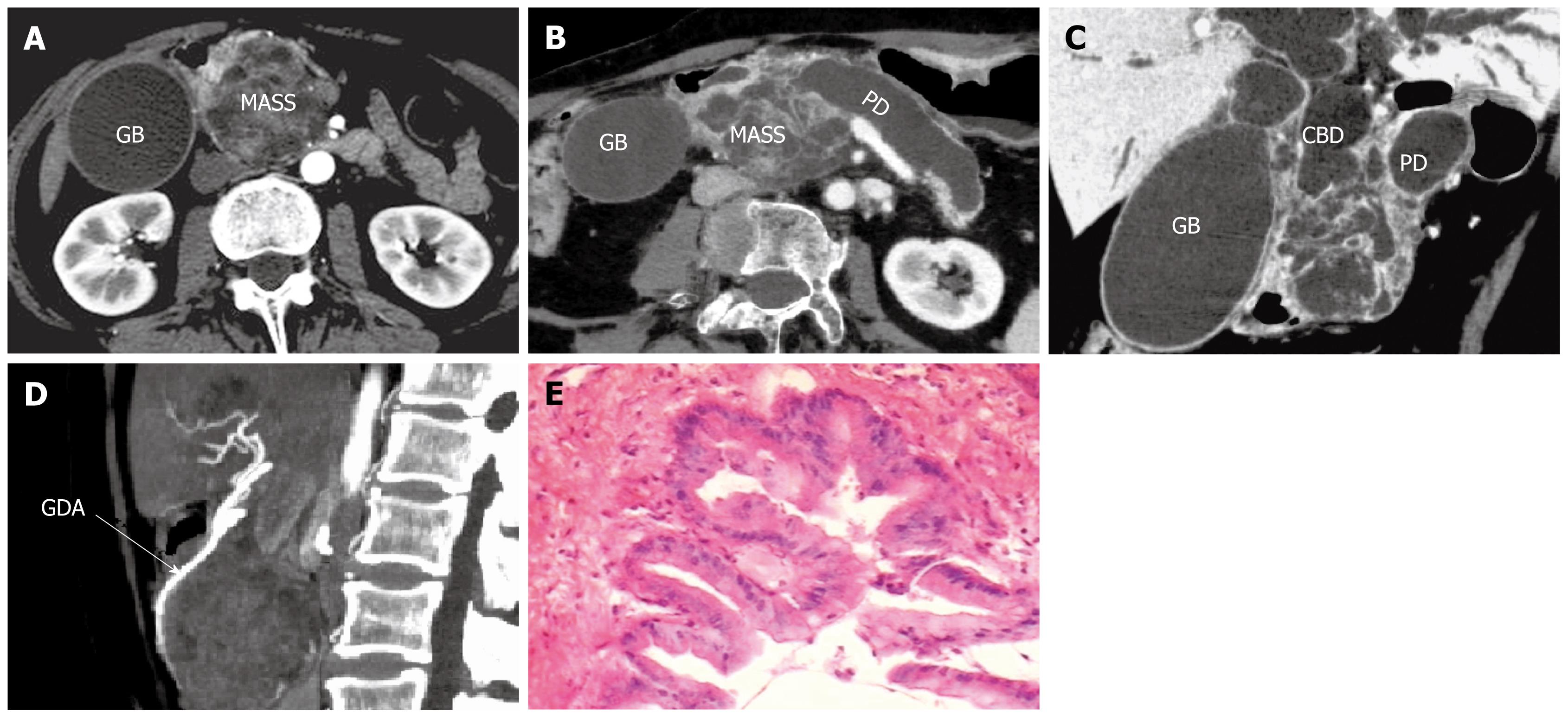
Figure 3 Pathologically confirmed malignant combined-type IPMN in a 65-year-old man with jaundice and abdominal pain for about 1 year.
A: An 8 cm cystic and solid mass (MASS) was seen in the axial arterio-phased MDCT image with contrast agents. The gallbladder (GB) was distended; B: The heterogeneous mass was shown with severe dilatation of the main pancreatic duct (PD) and the gallbladder (GB) in the CR image; C: The profile of the dilatation of pancreatobiliary system (CBD, common bile duct) was entirely depicted in the MPVR image; D: The gastroduodenal artery (GDA) showed irregularity as a result of infiltration of the tumor; E: The tumor consisted of papillary proliferations of tall columnar mucin-producing epithelium. Atypical epithelium characterized by enlarged nuclei (HE, × 150).
- Citation: Tan L, Zhao YE, Wang DB, Wang QB, Hu J, Chen KM, Deng XX. Imaging features of intraductal papillary mucinous neoplasms of the pancreas in multi-detector row computed tomography. World J Gastroenterol 2009; 15(32): 4037-4043
- URL: https://www.wjgnet.com/1007-9327/full/v15/i32/4037.htm
- DOI: https://dx.doi.org/10.3748/wjg.15.4037


