Copyright
©2009 The WJG Press and Baishideng.
World J Gastroenterol. Aug 28, 2009; 15(32): 4016-4022
Published online Aug 28, 2009. doi: 10.3748/wjg.15.4016
Published online Aug 28, 2009. doi: 10.3748/wjg.15.4016
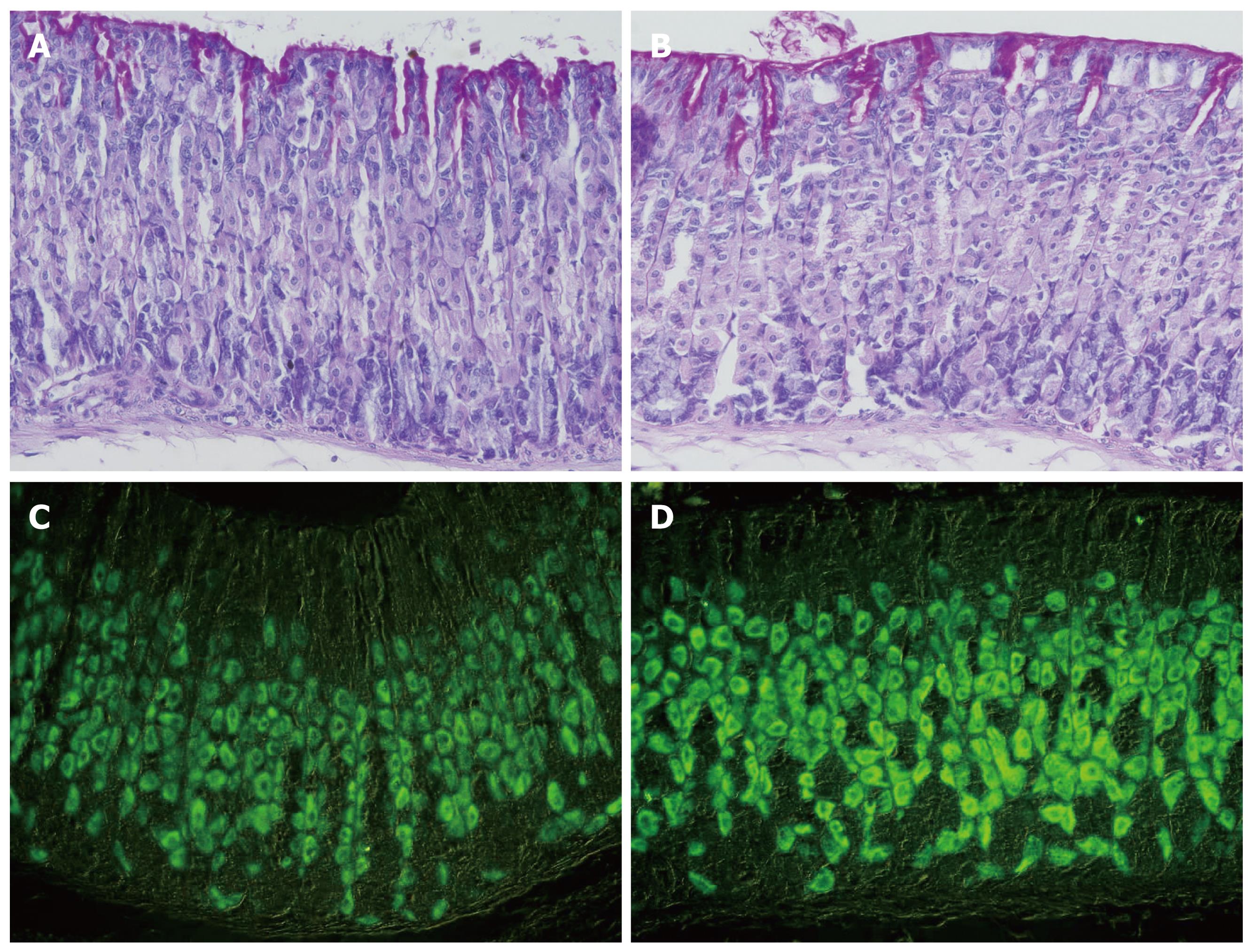
Figure 3 Gastric mucosal tissue sections of control (A, C) and smoke-treated (B, D) mice.
A and B demonstrate the gastric mucosae of control and treated mice stained with periodic acid-Schiff and hematoxylin. No difference is noted in parietal cells of control and treated tissues. Immunohistochemical labeling of parietal cells in the gastric mucosa of control (C) and smoke-treated (D) mice with antibodies specific for H,K-ATPase β-subunit. Labeled parietal cells are distributed throughout the gastric glands. Note the difference in the labeling intensity of parietal cells in control vs smoke-treated mice, × 400.
- Citation: Hammadi M, Adi M, John R, Khoder GA, Karam SM. Dysregulation of gastric H,K-ATPase by cigarette smoke extract. World J Gastroenterol 2009; 15(32): 4016-4022
- URL: https://www.wjgnet.com/1007-9327/full/v15/i32/4016.htm
- DOI: https://dx.doi.org/10.3748/wjg.15.4016


