Copyright
©2009 The WJG Press and Baishideng.
World J Gastroenterol. Aug 21, 2009; 15(31): 3950-3953
Published online Aug 21, 2009. doi: 10.3748/wjg.15.3950
Published online Aug 21, 2009. doi: 10.3748/wjg.15.3950
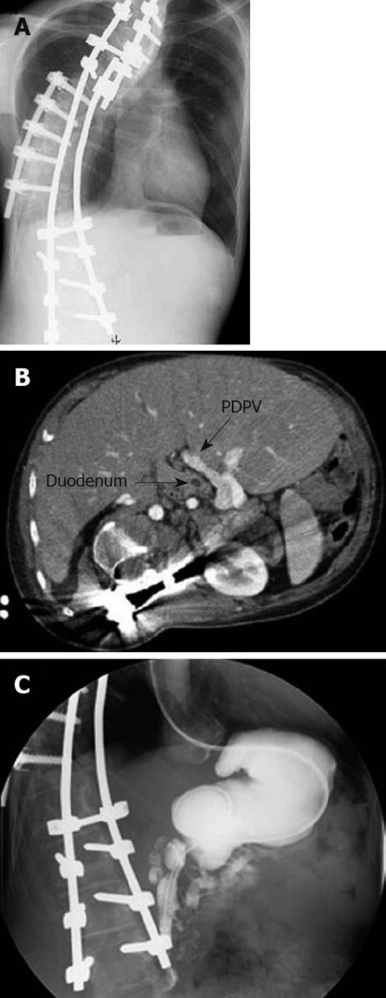
Figure 1 Images at admission.
A: Plain X-ray film. Although the spine was corrected by a previous orthopedic operation, a severe scoliosis with right side protrusion was found at admission; B: Abdominal CT scan. The preduodenal portal vein (PDPV) was found to be located in the anterior side of the duodenum; C: Contrast study of the upper gastrointestinal tract. A contrast study of the duodenum showed a stenosis ranging from the second to third portions. In addition, the contents flowed to both the dilated main pancreatic duct and common bile duct.
- Citation: Masumoto K, Teshiba R, Esumi G, Nagata K, Nakatsuji T, Nishimoto Y, Yamaguchi S, Sumitomo K, Taguchi T. Duodenal stenosis resulting from a preduodenal portal vein and an operation for scoliosis. World J Gastroenterol 2009; 15(31): 3950-3953
- URL: https://www.wjgnet.com/1007-9327/full/v15/i31/3950.htm
- DOI: https://dx.doi.org/10.3748/wjg.15.3950


