Copyright
©2009 The WJG Press and Baishideng.
World J Gastroenterol. Aug 21, 2009; 15(31): 3947-3949
Published online Aug 21, 2009. doi: 10.3748/wjg.15.3947
Published online Aug 21, 2009. doi: 10.3748/wjg.15.3947
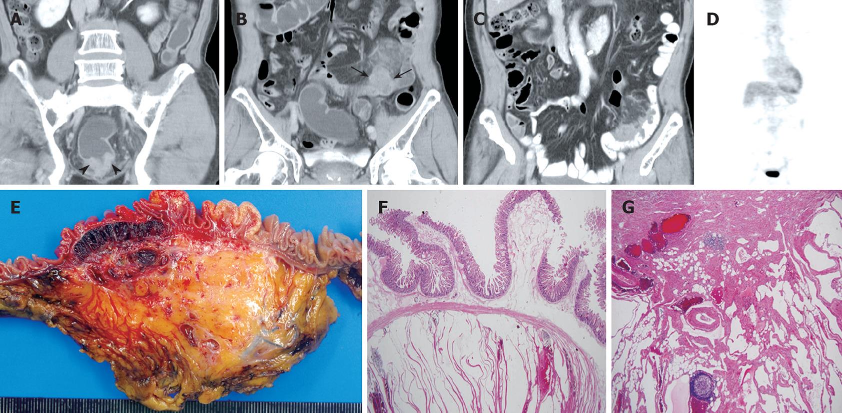
Figure 1 A 71-year-old man with rectal cancer and mesenteric lymphangioma.
A, B: Contrast-enhanced, coronal CT images show irregular and concentric rectal wall thickening (arrow heads), a nodular soft-tissue-density mass, and hazy strands in the jejunal mesentery (arrows); C: Follow-up, contrast-enhanced, coronal CT obtained one year after laparoscopic rectal cancer resection, reveals clear demarcation of the jejunal nodular lesions and infiltrative soft tissue masses in the adjacent mesentery. No remarkable jejunal obstruction was found; D: 18F-FDG PET shows no 18F-FDG uptake in the jejunum or mesenteric lesions; E: The cut section of the jejunum reveals a dark-red, multiloculated, cystic lesion measuring 8.0 cm × 5.0 cm in the mucosa to the subserosa; F, G: Histopathologic view of the tumor shows numerous, multiloculated, cystically dilated lymphatic spaces lined by attenuated endothelium in the entire jejunal wall (HE stain, × 40) and adjacent mesentery (HE, × 20).
- Citation: Hwang SS, Choi HJ, Park SY. Cavernous mesenteric lymphangiomatosis mimicking metastasis in a patient with rectal cancer: A case report. World J Gastroenterol 2009; 15(31): 3947-3949
- URL: https://www.wjgnet.com/1007-9327/full/v15/i31/3947.htm
- DOI: https://dx.doi.org/10.3748/wjg.15.3947


