Copyright
©2009 The WJG Press and Baishideng.
World J Gastroenterol. Aug 21, 2009; 15(31): 3874-3883
Published online Aug 21, 2009. doi: 10.3748/wjg.15.3874
Published online Aug 21, 2009. doi: 10.3748/wjg.15.3874
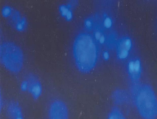
Figure 4 Morphological changes of MGC803 cells 24 h after treatment with 30 μmol/L 15d-PGJ2.
Cell shapes were observed by fluorescence microscopy (Hoechst 33342 staining, × 400). Nuclear chromatin condensation, marginalization of nuclear chromatin and half moon formation, nuclear fragments with bright chromatin and apoptotic bodies were easily identified in some cells.
- Citation: Ma XM, Yu H, Huai N. Peroxisome proliferator-activated receptor-γ is essential in the pathogenesis of gastric carcinoma. World J Gastroenterol 2009; 15(31): 3874-3883
- URL: https://www.wjgnet.com/1007-9327/full/v15/i31/3874.htm
- DOI: https://dx.doi.org/10.3748/wjg.15.3874


