Copyright
©2009 The WJG Press and Baishideng.
World J Gastroenterol. Aug 21, 2009; 15(31): 3865-3873
Published online Aug 21, 2009. doi: 10.3748/wjg.15.3865
Published online Aug 21, 2009. doi: 10.3748/wjg.15.3865
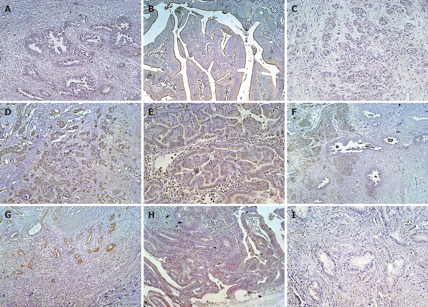
Figure 3 LNβ3 expression in biliary tract cancer tissues.
A-C: Extrahepatic bile duct cancer tissues; A: Well differentiated tubular adenocarcinoma positive for invasive front dominant staining; B: Papillary adenocarcinoma positive for diffuse staining; C: Moderately differentiated tubular adenocarcinoma negative for staining; D-F: Gallbladder cancer tissues; A-C: Extrahepatic bile duct cancer tissues; D: Moderately differentiated tubular adenocarcinoma positive for invasive front dominant staining; E: Papillary adenocarcinoma positive for diffuse staining; G-I: Carcinoma tissues of the ampulla of Vater; G: Well differentiated tubular adenocarcinoma positive for invasive front dominant staining; H: Well differentiated tubular adenocarcinoma positive for diffuse staining; I: Well differentiated tubular adenocarcinoma negative for staining.
- Citation: Oka T, Yamamoto H, Sasaki S, Ii M, Hizaki K, Taniguchi H, Adachi Y, Imai K, Shinomura Y. Overexpression of β3/γ2 chains of laminin-5 and MMP7 in biliary cancer. World J Gastroenterol 2009; 15(31): 3865-3873
- URL: https://www.wjgnet.com/1007-9327/full/v15/i31/3865.htm
- DOI: https://dx.doi.org/10.3748/wjg.15.3865


