Copyright
©2009 The WJG Press and Baishideng.
World J Gastroenterol. Aug 14, 2009; 15(30): 3748-3756
Published online Aug 14, 2009. doi: 10.3748/wjg.15.3748
Published online Aug 14, 2009. doi: 10.3748/wjg.15.3748
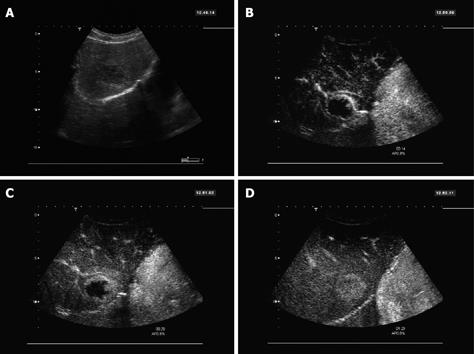
Figure 2 36-year-old female.
A: 4.0 cm hypoechoic lesion in segment 8 found in B-Mode sonography; B: Contrast-enhanced sonography with SonoVue® showed a peripheral enhancement with nodular contrast accumulations 14 s after injection; C: Slow progression of the enhancement from the periphery towards the center of the lesion, with a broader peripheral enhancement zone seen at 20 s; D: After 1.5 min, the lesion is completely filled with contrast and appears hyperenhanced compared to the surrounding normal liver tissue. The enhancement pattern is typical for a hemangioma.
- Citation: Trillaud H, Bruel JM, Valette PJ, Vilgrain V, Schmutz G, Oyen R, Jakubowski W, Danes J, Valek V, Greis C. Characterization of focal liver lesions with SonoVue®-enhanced sonography: International multicenter-study in comparison to CT and MRI. World J Gastroenterol 2009; 15(30): 3748-3756
- URL: https://www.wjgnet.com/1007-9327/full/v15/i30/3748.htm
- DOI: https://dx.doi.org/10.3748/wjg.15.3748


